

Musculoskeletal System
- Created by: Charlotte170289
- Created on: 04-12-20 14:45
Components of the Musculoskeletal System

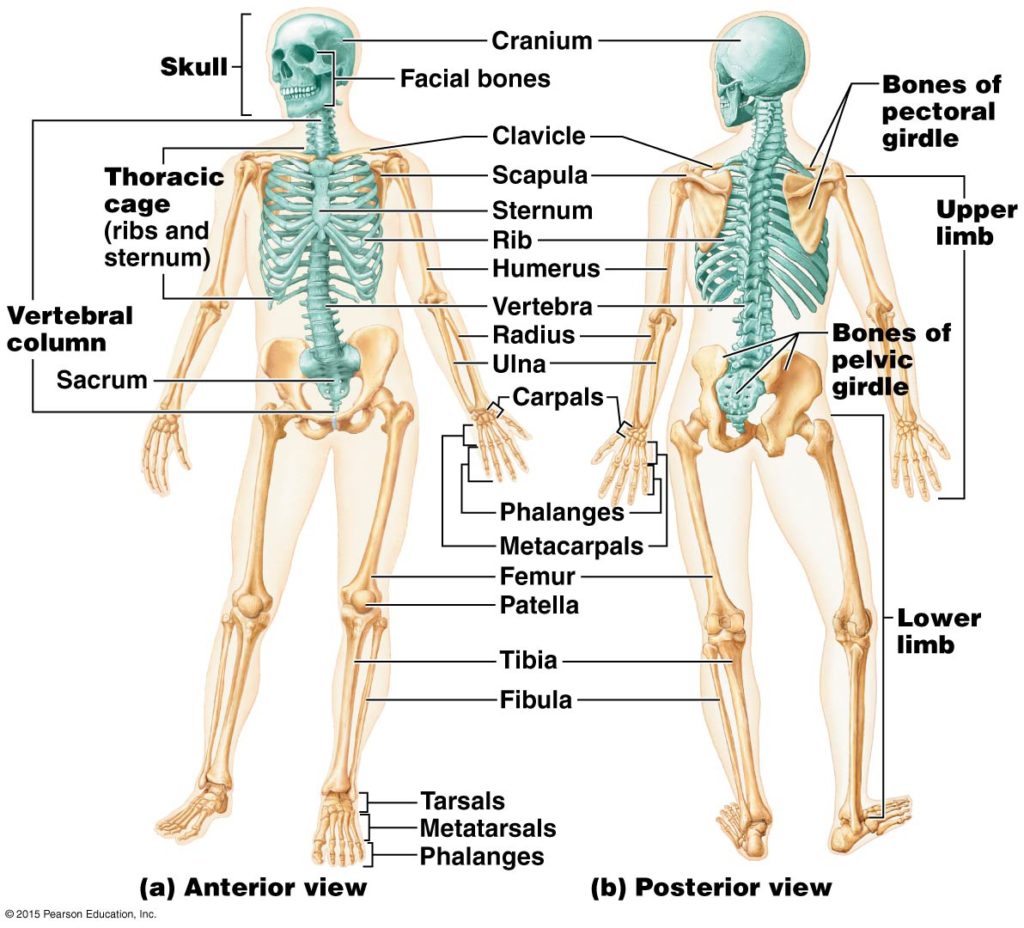
The skeleton- Axial skeleton
Axial skeleton:
- Is formed bu the vertebral column (the spine), rib cage and the skull
- The upright posture of the human body is maintained by the axial skeleton, which transmits the weight from the head, the truck (torse) and the upper extremities down to the lower extremities at the hip joints

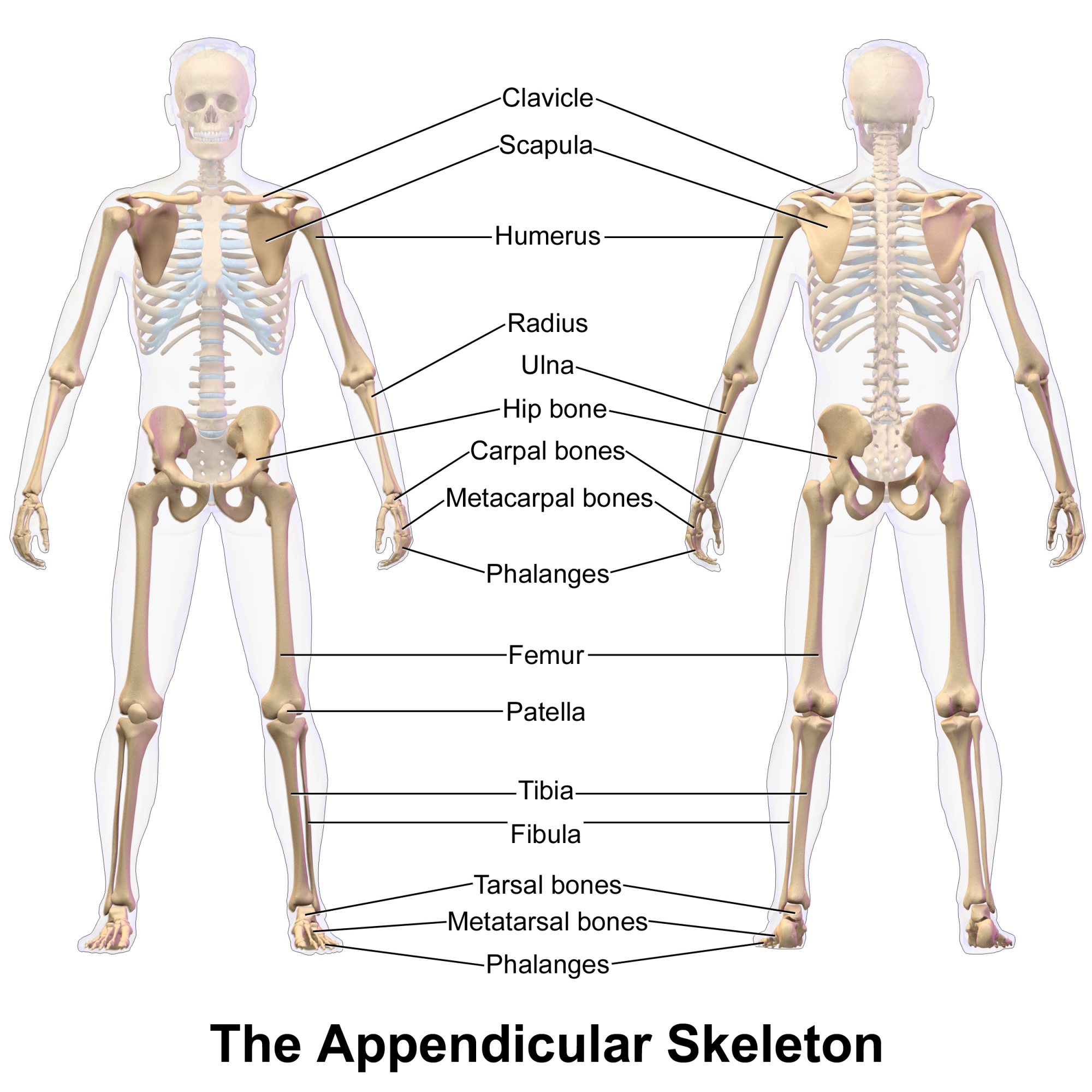
The skeleton- Appendicular skeleton
Appendicular skeleton:
- Is made up of the pelvic, pectoral (shoulder) gridles and the bones and cartilages of the upper and lower limbs (arms and legs)
- Their functions are to make movement possible and to protect the major organs of the digestive, excretion and reproduction systems
The skeleton
Classification of bones
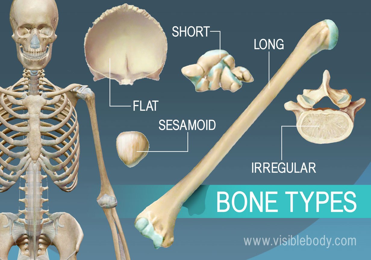
Classification of bones- Sesamoid bones
Sesamoid bones:
- Are usually short ot irregulat bones embedded in a tendon.
- The most obvious example of this is the Patella which sits within the Patella tendon.
- Other sesamoid bones are the Pisiform (smallest Carpal) and the two small bones at the base of the 1st metatarsal.
- Sesamoid bones are usually present in a tendon where it passes over a joint which serves to protect the tendon.


Classification of bones- Irregular bones
Irregular bones:
- Are bones in the body that do not fall into any other category due to their non-uniform shape.
- Some examples of irregular bones are: the vertebrae, sacrum and mandible.
- They primarily consist of cancellous bone with a thin outer layer of compact bone.

Classification of bones

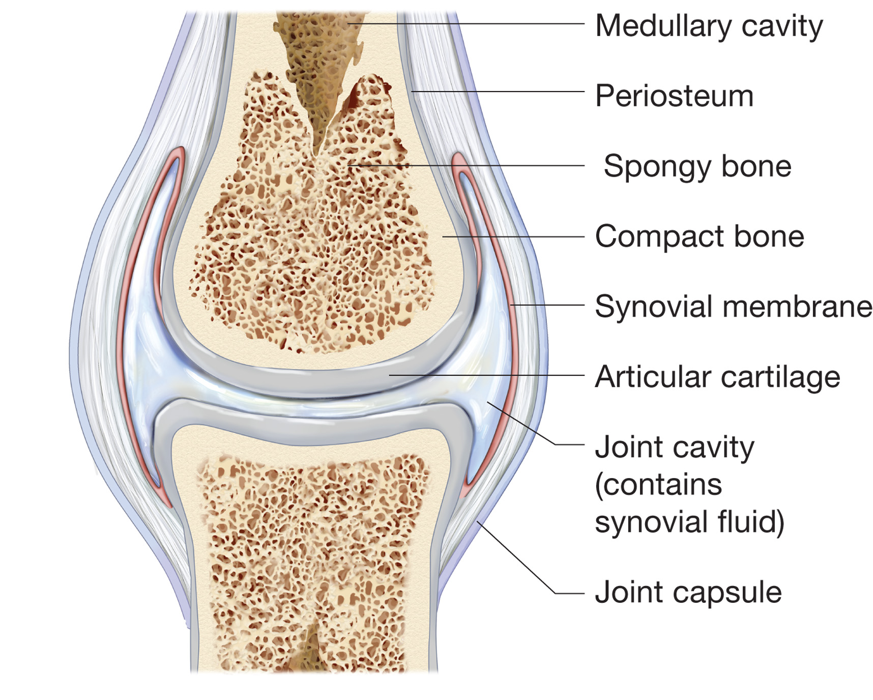
Bone structure
Bones are:
- Covered by a layer of protective periosteum
- The outer layer is compact bone
- The inner layer is a spongy bone
- Hollow marrow cavity- Yellow marrow adipose (fat) tissue- Red marrow (oly in epiphyses and flat bones in adults)
Compact bone structure
The Haversian system
- Also known as an Osteon
- It is a series of concentric circles called lamellae found in the compact bone.
- Down the middle of each of these systems is a hollow tube that holds a blood vessel.
- Within the Lamellae of each Haversian system are a series of spaces called Lacunae. The Lacunae holds the Osteocytes or bone cells.
- The blood vessels that run down the middle of the Haversian system provides nutrients to the living bone tissue
- Nerves and Lymph vessels are also found in the Haversian canals.

Vertebral column
- It is formed from individual bones called vertebrae which houses the spinal canal that encloses and protects the spinal cord.
- Vertebrae are defined by the regions of the vertebral column that they occur in
- It usually consists of 33 vertebrae: the upper 24 are articulating vertebrae, separated by intervertebral disks and the lower nine are fused, five fused in the sacrum and four in the coccyx.

Joints
There are three structural classifications of joints:
- Fibrous joint- joined by dense regular connective tissue that is rich in collagen fibres e.g. Skull
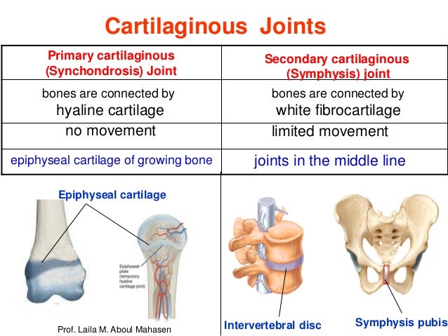
- Cartilaginous joint- are formed by a pad of fibrocartilage, a tough material that acts as a shock absorber e.g. pubic symphysis
- Synovial joint- not directly joined- the bones have a synovial cavity and are united by the dense irregular connective tissue that forms the articular capsule. Highly moveable found at the end of the long bone e.g. knee. They have a space/cavity between the articulating bones. The end of the bones are held together by a sleeve of fibrous tissue and the capsule are lubricated with a small amount of fluid.
Primary and Secondary cartilaginous joints
Synovial joint
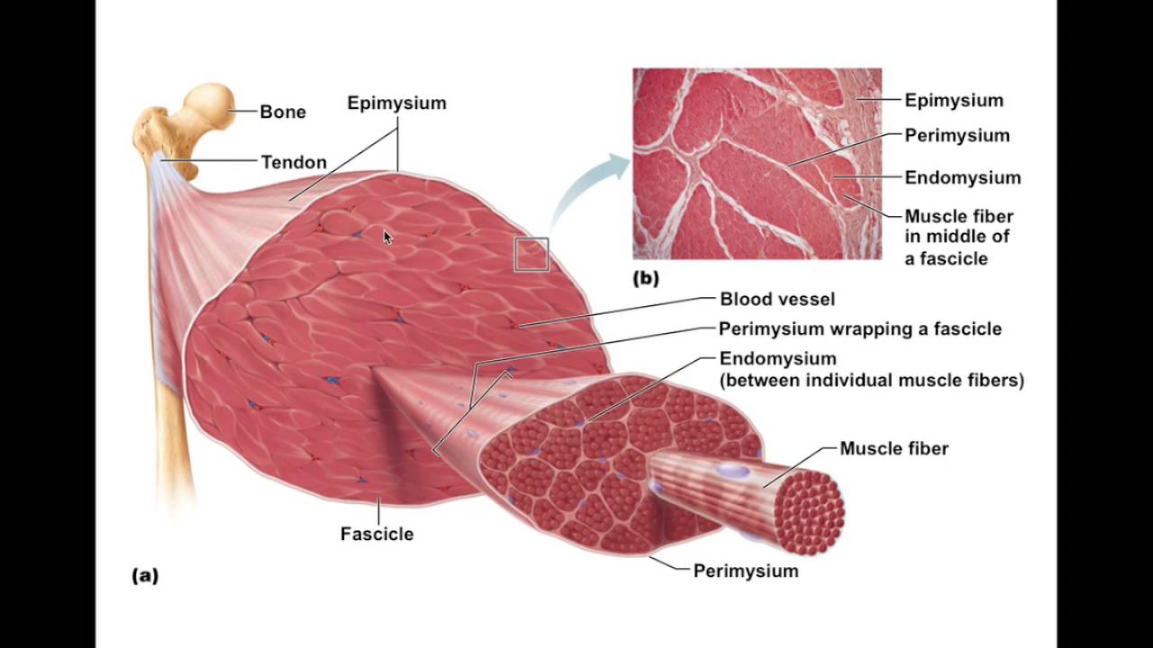
Skeletal muscle
- Connective tissues cover the muscle fibres (endomysium), bundles of cells (perimysium) and the muscle body.
- Connective tissues come together at the muscle ends to form tendons
Muscle contraction (shortening)
- The skeletal muscle cells contracts in response to stimulation from a nerve fibre which supplies the muscle cell usually around have way along its length.
- Nerve impulses from the brain stimulate muscle cells in the muscle body e.g. biceps brachii
- When the action spreads down the nerve along the sarcolemma, it conducts deep within the muscle cell through a special network of channels that run through the sarcoplasm, releasing calcium from the intracellular stores
- Calcium triggers the binding of myosin to the actin filament next to it, forming a cross bridge
- Protein filaments (ATP) then provides the energy for the 2 filaments to slide over each other, pulling the z lines at each end of the sarcomere closer to one another, shortening the sarcomere
- If enough fibres are stimulated the whole muscle with contract (shorten).
- The Muscles pulls on tendons creating leverage on the bones enabling movement

Movement
- Mucles work in pairs or groups called antagonistic pairs- many muscles/ muscle groups of the body are arranged so that thir actions oppose one another.
- In order to move a body part the muscles or its tendons must stretch across at least one joint
- When it contracts the muscle pulls one bone to another, the action caused by contraction of the biceps brachii is flexion
- The action caused by contraction of the Triceps brachii is extension

Smooth muscle

Comments
No comments have yet been made