Nerve and Muscle Physiology

?
  • Created by: rosieevie
  • Created on: 19-01-17 12:34

Divisions of the Nervous System

CNS:

  • Brain
  • Spinal Chord

PNS:

  • Sensory divison - detects environmental changes
  • Motor division
    • Somatic - voluntary
    • Autonomic - involuntary
      • Sympathetic - increases to norm
      • Parasympathetic - decreases to norm
1 of 20

Spinal Cord and PNS

Grey matter - cell neuron bodies

White matter - fibre tracks (milanated axons)

Dorsal root ganglion - cluster of nerve cell bodies outside CNS

Dorsal root - where afferent fibres enter spinal chord

Ventral root - where efferent fibres leave spinal chord

Afferent fibre - carry signals to CNS (sensory info)

Efferent fibre - carry signals from CNS to PNS (motor info)

Nucleus - nerve cell body cluster within CNS

Reflex - predictable stereotypical response to specific stimulus

Renshaw cells - inhibatory cells stopping other neurones becoming excited (lateral inhibiton)

2 of 20

Monosynaptic Neural Simple Reflex Circuit

  • Extensor muscle stretched and muscle spindle stimulated
  • Primary afferent neuron excited
  • Synapse
  • Efferent alpha motor neuron stimulated
  • Extensor muscle stimulated and contracts
  • Flexor muscle relaxes

1 synapse, 2 neurones

3 of 20

Autonomic Motor Division - Involuntary

(http://hal.bim.msu.edu/ome2/Content/Autonomic/Overview/Images/NeuroTrans.jpg)

  • Sympathetic - stress
  • Parasympathetic - resting conditions
4 of 20

Sympathetic and Parasympathetic Nervous System

  • Effect always have at least 1 synapse in outside CNS ganglion
  • Preganglionic efferent axons myelinated
  • Chloinergic ganglionic synapses - acetylcholine
  • Postganglionic non-myelinated
  • Postganglioninc neurones form plexi (spinkler system) w/ many synapses
5 of 20

Neurone

(http://teachmeanatomy.info/wp-content/uploads/Structure-of-a-Neurone.jpg)

  • Dendrite - branches recieving signals, contain postsynaptic membrane
  • Soma - cell body containing nucleus and organelles
  • Axon - output process, different extensions depending on neurone
  • Boutons - swellings at axon terminals contain pre-synaptic memebrane
  • Myelin Sheath - insulating layer to quickly conduct impulses (Schwann cells - glial)
  • Nodes of Ranvier - gaps where action potential occurs
6 of 20

Exciting a Neurone

Resting membrane potential - high energy state

Outside = 0mV

Inside = -60mV (negative)

  • More Na+ outside 
  • More K+ inside - leak out of cell (selective permeability to K+ ions)
  • More Cl- inside

= CONCENTRATION GRADIENT (maintained by ion pumps against gradient e.g. Na+/K+ ATPase)

Electrogenic ion pump - pump unequal number of ions (3Na+,2K+) generates a electrical gradient

Increase concentration gradient -> K+ ions move down out fo cells = more -ve

7 of 20

Electrochemical Gradients

Electrochemical gradient - how far equillibrium potential is from resting membrane potential

Ions move down electrochemical gradient

Equilibrium - electrical gradient balances concentration gradient

Equilibrium potential (mV) - potential difference between 2 membrane sides

EmV = 61.5 log([K+out]/[K+in])            NERNST EQUATION

When the membrane = Emv there is no net flux of ions

8 of 20

Hodgkin and Huxley

Squid axon tested to see if EmV determined soley by K+ permeability

Should be straight correlation if just K+ ions

However - devaiation

MEMBRANE AT REST IS SLIGHTLY PERMEABLE TO Na+ - set by tansmembrane ion channels

Em = -60mv to -70mv

9 of 20

Goldman Constant Field Equation

(http://clinicalgate.com/wp-content/uploads/2015/05/B9780443103216000060_f006-005-9780443103216.jpg)

60 should be 61.15

10 of 20

Ions

 ECl<Em (more negative) due to ion exchangers -> increased Cl- hyperpolarises neurones

At rest Na+ ins not at equilibrium = high energy state

11 of 20

Stimulations

Weak Electrical Stimulaton

Electrotonic conduction - stimulus passively decreases in amplitde

Strong Electrical Stimulation

Generates action potential - propogates along axon

All or nothing response = axon havs threshold for activation

12 of 20

Action Potential

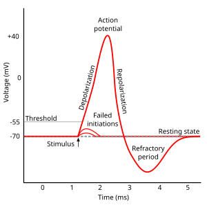
Measured using voltage-clamp technique.
Action potential spike due to transient increase in Na+ permeability (voltage-gated Na+ channels open) due to membrane depolarisation

13 of 20

Action Potential Explained

  • Resting potential - PK>>PNa
  • Passive depolarisation - no change in ionic permeability PK>>PNa
  • Threshold potential - voltage-gated Na+ channels activated, Na+ current depolarises membrane PNa>>PK -> Em gets closer to ENa
  • Postive feedaback cycle - more Na+ channels open
  • Em overshoots 0
  • Em approaches ENa (~+60mV) = current driving force decreases
  • Na+ channels inactivate - time-dependent inactivation gate
  • Delayed recifer K+ channels open, PNa decreases + PK increases re/hyperpolarisation
  • Em returns to resting value but undershoots
  • Both leaked and delayed K+ channels open - PK>>PNa = refractory period
  • Both Na+ channels and delayed rectifier channels deactivate - Em returns resting value
14 of 20

Refractory periods

Absolute refractory period - inexcitable membrane as all Na+ channels inactivated

Refractory period - activation threshold is increase as some Na+ channels at resting state

15 of 20

Action Potentials are very Efficient

After adding a neurotoxin to nerve axon action potentials still conducted for considerable period of time after

Only a few ions are moved to generate action potentials as membrane is a capacitor - seperates and stores electical charge (around 1x10-12mol Na+ needed)

COMPOUND ACTION POTENTIAL - the sum of the action potentials in a bundle of axons 

Compound action potential amplitude = how many axons excited and strength of stimulus

  • Increases as stimulus increase until all axons in nerve are excited/recruited
16 of 20

Myelinated Neurones

Increases conduction velocity - increases out resistence:in resistence

  • Peripheral nerves - Schwann cells
  • CNS - oligodendrocytes

Unmyelinated axons:

  • Adjacent regions exchange charge - depolarises next section
  • Unidirectional
  • Velocity ~5m/sec
  • Increasing axon diameter increases velocity

Myelinated axons:

  • Salatory conduction - AP skips between nodes of Ranvier
  • Uniderctional
  • Zero sigbal loss 
  • Small axon diameters
  • Rapid transmission ~100m/sec
  • Voltage-gated channgels at nodes of Ranvier
17 of 20

Ca2+ Dependence of Synapses

Ca2+ dependence of synaptic transmission

Possible to block Na+ chanels and record postsynaptic response = Na+ isn't cause

Removing extracellular Ca2+ abolished nerurotransmitter release.

18 of 20

Synapses

  • Neurotransmitter synthesised and stored in vesicles
  • Action potential at presynaptic bouton = depolarisation
  • Voltage-gated Ca2+ channels open = Ca2+ into presynaptic bouton
  • Vesicles fuse with presynaptic membrane
  • Neurotransmitter released into synaptic cleft by exocytosis
  • Neurotransmitter binds to receptor molecules
  • Postsynaptic channels open/close
  • Postsynaptic current causes excitatory or inhibitory potential that changes cell excitability
  • Neurotransmitter deactivated by reuptake or enzymatic degredation
19 of 20

Excitatory/Inhibitory Postsynaptic Currents/Potent

Excitatory

Current dspolarizes postsynaptic membrane = potentials summate until threshold reached for action potential generation

Inhibatory Currents

Current flow through inhibatory neurotransmitter activated channels

Inhibatory Potentials

Summate to reduce excitability (hyperpolarise cells), reduce probability of action potential generation

20 of 20

Comments

bradhasaids

Report

just WOW just WOW man

Similar zoology resources:

See all zoology resources »See all Action Potentials resources »