2.1.1 Cell structure
- Created by: Maria
- Created on: 06-07-18 22:32
(a) the use of microscopy to observe and investigate different types of cell and cell structure in a range of eukaryotic organisms
To include an appreciation of the images produced by a range of microscopes; light microscope, transmission electron microscope, scanning electron microscope and laser scanning confocal microscope.
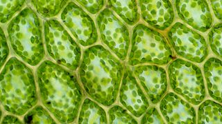
Light (optical) microscope
Magnification = 1500x
Resolution = 200nm
Advantages
- Cheap
- Easy to use
- Portable
- Can study living organisms
Disadvantages
- Low resolution gives no detail
- Non-coloured specimens must be stained
- Low magnification
Electron microscopes
Resolution = 0.1nm (2000x more than light microscope)
Specimen preparation:
- Dehydrated
- Stained
- Sectioned
Advantages
- Detailed images by high resolution
- High magnification
Disadvantages
- Cells must be dead
- Expensive
- Need training to use
- Electron beam damages tissue
- Time-consuming
-Transmission electron
Magnification = 500,000x Emits an electron through the sample, denser parts appear darker giving contrast to the 2D image
-Scanning electron
Magnification = 100,000x Emits an electron beam into the sample, which bounces off the sample and is received on a sensor, producing a 3D image.
Can add colour
Laser scanning confocal microscope
High resolution
Advantages
- Can create a 3D image
- Clearer images than a light microscope
- Used in bioresearch
Disadvantages
- Need skill and training to use

(b) The preparation and examination of microscope slides for use in light microscopy
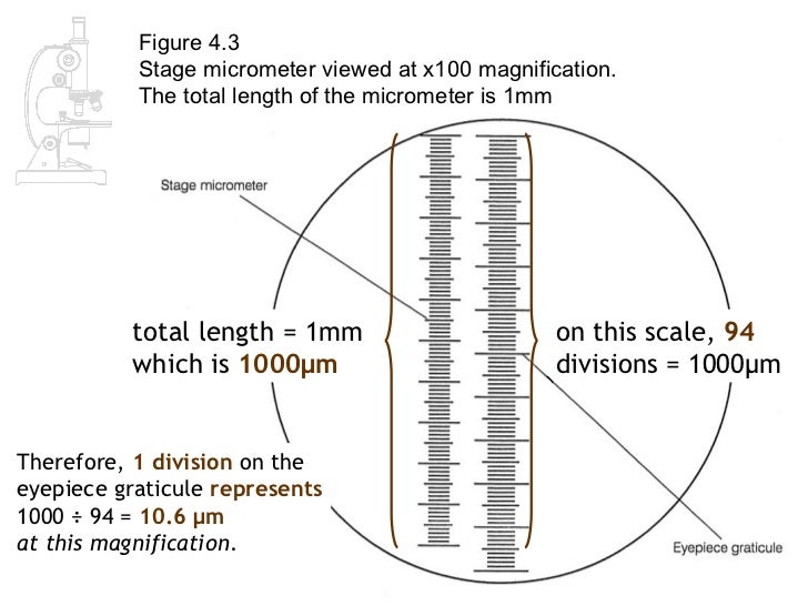
Including the use of an eyepiece graticule and stage micrometre
You need to calibrate the eyepiece graticule using the stage micrometre to allow you to measure the size of the cells.
- Align the stage micrometre (a microscope slide with a scale on it) with the eyepiece graticule. Then use the reading from the scales to calculate the calibration factor from the objective lens
Centimetre (cm) x10
= Millimetre (mm) x1000
= Micrometre (μm) x1000
= Nanometre (nm)
(c) the use of staining in light microscopy
To include the use of differential staining to identify different cellular components and cell types.
Some biological matter is not coloured, making it difficult to see. Staining can be used where it binds to specific parts
- Acetic stains DNA dark red
- Eosin stains cytoplasm
- Sudan red stains lipids
- Iodine stains cellulose yellow and starch blue/black or violet
- Methylene blue stains DNA, RNA
(d) the representation of cell structure as seen user the light microscope using drawings and annotated diagrams of whole cells or cells in sections of tissue
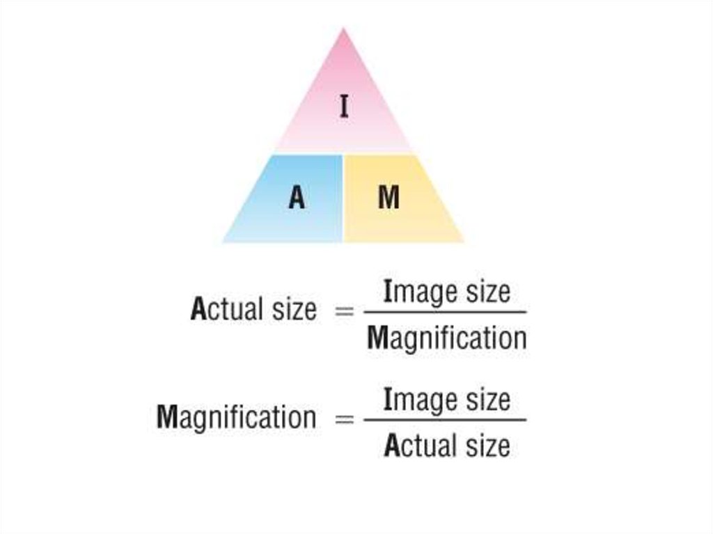
(e) the use and manipulation of the magnification formula
magnification = image size / object size
(f) the difference between magnification and resolution
To include an appreciation of the differences in resolution…
Comments
No comments have yet been made