Applied Science: Unit 1 Biology (including cells, tissues, microscopy etc)
- Created by: x.Becca.x
- Created on: 26-01-20 11:26
Animal Cells
Animal cells carry out funtions such as making proteins, replicating DNA and storing energy. They have the same organelles as plant cells that perform the same functions:
-Nucleas and Nucleolus, Cell Wall and Cell Membrane, Ribosomes and Mitochondria, Golgi Apparatus, Rough ER/ Smooth ER, Cytoplasm
However, they do not have:
-Vacuole or Chloroplasts
Gram Staining
Gram staining is a test to show which type of bacteria you have. You dye the cells with safranin (a pink colour) and crystal violet (purple colour).
-If a cell turns pink, it means that it is a gram negative. This means the cell was unable to retain the purple dye. The cell wall of a gram negative bacteria is made up of a thick peptidoglycan layer with no lipid membranes. Gram negative bacteria are hard to get rid of- more anti-biotic resistant.
-However, if a cell turns purple, it means that it is a gram positive. This is because the cell wll is made up of a thinner layer of peptidoglycan and has lipid membranes. Therefore, it was able to wash of the crystal violet purple stain and retain the pink safranin stain.


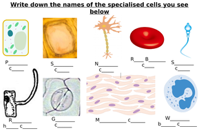
Specialised Cells (2)
Pallisade Mesophyll- Found in leafs on plant cells. These help the plant phtosynthesize as they absorb light. It consists of chlorplasts and chlorophyll making the green colour.
Xylem- Transports water, nutrients and minerals upwards a plant from the roots. Long tubes are used to trasnport this material.
Phloem- A vascular tissue in charge of transport of materials and distrubution of organic nutrients. It is a pathway to signaling molecules. They help keep the plant upright.
White blood cell- A cellular component of blood. It has a circular shape. It lacks haemoglobin but has a nucleus. It defends the body against disease and infection by using pathogens and producing antibodies. This process can be called phagocytosis.
Microscopy
Microscopy= Using a microscope to see something that is too small too see with the naked eye.
There are two types of microscopes: An electron microscope and a light microscope.
You can calculate the magnification of an image:
 image size= actual size x magnification.
Tissues
Tissue: A group of similar cells that work together to carry out a particular function. Tissues work together to form organs. There are different types of tissues within our bodies. The main types of tissues are: epithelial and endothelial.
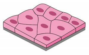
Epithelial: These tissues cover internal and external body surfaces and secreting organs. There are different types of epithelial tissue: squamous, collumnar and cubodial. There are also three ways to describe these cell layers: simple, stratified and psuedo-stratified.
Squamous epithelial tissue: They are a barrier between the under layers and exterior environment. They provide protections. They are one layer thick made of sqaumous cells. These cells are large, thin and flat and contain a rounded nucleus. These can be found in the alveoli of the lungs and the capillaries. This is the simple type.There can also be stratified type of this tissue. Smoking causes inflammationb in the lungs and damages this type of tissue.
Tissues (2)
Collumnar epithelial tissue: These cells are elongated and column-shaped. These are usually located near the base of cells. The nuclei is also elongated. They from the lining of the stomacha nd intestines. It's functions inckude absorption and secretion. This tissues have cillia (hair-like structures) that sweepaway pathogens. This is the pseudo-stratified type, it can also be simple- which has much shorter cells than the picture.

This type of tissue can secrete mucus nad trap unwanted partciles. Therefore protecting the lungs from harmful bacteria.
Tissues (3)
Cuboidal epithelial tissue- This tissue is a single cell layer thick and is made up of cube shaped cells. This type of tissue is found lining parts of the body such as kidney tubules and respiratory bronchioles. It is a protective lining to organs in the body. This is the simple type of tissue. It can also be stratified cuboidal tissue.
Endothelial Tissue
Endothelial tissue- the endothelium refers to the lining of the interior surface of blood vessels and lymphatic vessels. Itcirculates blood or lymph in the lumen and the rest of the vessel wall. They align and elongate fluid flow.

Synaptic Transmission
Neurotrasmitters are chemicals held in synaptic vesicles which are stored inside the presynaptic neurone. The calcium channels open, releasing stored calcium ions. Using these calcium ions, the neurotrasmitters move and diffuse out of the pre-synaptic neurone, through the synaptic cleft and into the receptors on the post-synaptic neurone. From here, the neurotransmitters are absorbed into the post-synaptic neurone to create an electrical impulse. Examples of some neurotransmiotters would include: Acetylcholine, Dopamine, Glutamate, Glycine, Carbon monoxide and Seretonin.
![]()
Nervous Tissue
Resting potential- is given to a neurone at rest.
Action potential- happens when a neurone sends information/ an impulse to another neurone through the spine of brain. Part of the neural membrane opens to allow positivley charged ions in and negativle charged ions out.
Nodes of Ranvier-They gap between schwann cells covered in a myelinated sheath. The information jumps from node to node to create a impulse.

Comments
No comments have yet been made