infection of collateral cartilages, swelling/discharge from coronary band. tx by sx debridement - care DIPJ
11 of 17
What is a keratoma?
benign tumour of the hoof/solar horn. Intermittent lameness/discharge
12 of 17
How would you diagnose and treat a keratoma?
circular area of abnormal keratonisation within a discharging tract. smooth radiolucent defect in P3 on x ray. Sx to resect
13 of 17
What is canker?
chronic hypertrophy of the germinal layer of the frog epithelium. Fusobacterium/bacterioides. Dyskeratosis. Abnormal hyperkeratotic horn with keratolysis and fronds of unconnected intertubular horn
14 of 17
How would you treat canker?
mild - improve environment, debride, metronidazole bandages and abs, astringents, dilute formaline. Sx debridement if more advanced, bandage/shoe
15 of 17
What is white line disease?
Progressive crumbling, poor quality hoof wall with separation at white line
16 of 17
How would you treat white line disease?
renive abnormal horn, support remaining horse whith bar shoe/clips/acrylic, preven - correct environment, iodine feed supplementst
17 of 17
Other cards in this set
Card 2
Front
How would you treat a hoof crack?
Back
debride necrotic material, filler (Wire/plate) to stabilise, trim ,unload crack/bar shoe/quarter clips, tx underlying cause, abs
Card 3
Front
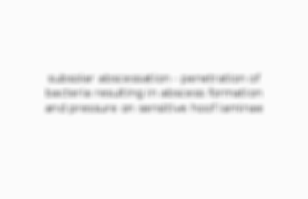
Which structures may be involved in an avulsion to the coronary band and hoof wall?
Back
Card 4
Front
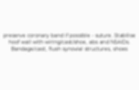
How would you treat an injury to the coronary band/hoof wall?
Back
Card 5
Front
What condition of the foot would you expect with increased digital pulse/hoof temperature and increased sensitivity to hoof testers?

Comments
No comments have yet been made