Electrical Activity of the Heart
5.0 / 5 based on 1 rating
- Created by: LBCW0502
- Created on: 13-02-19 17:02
Outline the conduction pathway in the heart (1)
SAN, atrial muscle depolarisation, AVN, bundle of His down septum, down to Purkinje fibres, apex of heart, depolarisation of ventricles. Conduction in AVN is the slowest (0.05 m/s). Conduction faster in fibres (Bundle of His/Purkinje)
1 of 58
Outline the conduction pathway in the heart (2)
Delay at AVN to ensure atrial empties and ventricles fill with blood. Contraction begins at apex (from bottom to top, ensures blood is squeezed out of ventricles into conductance vessels)
2 of 58
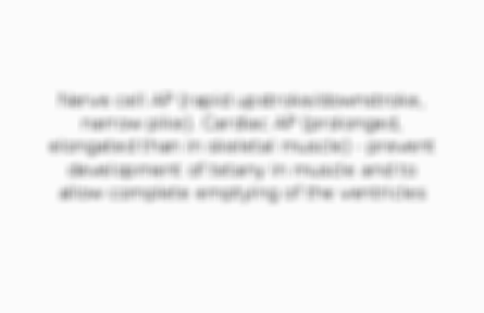
Describe features of the cardiac AP
Nerve cell AP (rapid upstroke/downstroke, narrow pike). Cardiac AP (prolonged, elongated than in skeletal muscle) - prevent development of tetany in muscle and to allow complete emptying of the ventricles
3 of 58
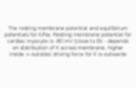
What is the voltage boundaries of the AP determined by? (1)
The resting membrane potential and equilibrium potentials for K/Na. Resting membrane potential for cardiac myocyte is -80 mV (close to Ek - depends on distribution of K across membrane, higher inside > outside) driving force for K is outwards
4 of 58
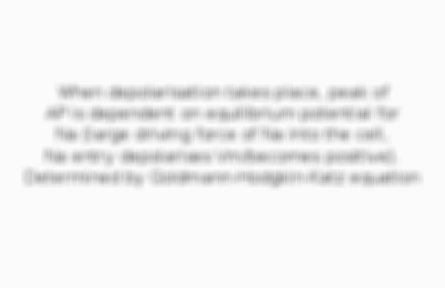
What is the voltage boundaries of the AP determined by? (2)
When depolarisation takes place, peak of AP is dependent on equilibrium potential for Na (large driving force of Na into the cell, Na entry depolarises Vm/becomes positive). Determined by Goldmann-Hodgkin-Katz equation
5 of 58
What happens during depolarisation? (1)
Initial rapid depolarisation results from a positive feedback at Na channels. Small depolarisation, few Na channels open, more depolarisation, more Na channels open, still faster depolarisation. Rapid upstroke of AP due to opening of Na channels
6 of 58
What happens during depolarisation? (2)
Gradual depolarisation towards threshold, to generate fast phase of AP (co-operative depolarisation)
7 of 58
What are the states of the sodium channels? (1)
Open state changes to inactive state (depends on time), during repolarisation it goes to closed state (states determined by conformational changes in the protein). Depolarisation only occurs when Na channels are in closed state
8 of 58
What are the states of the sodium channels? (2)
When inactive there is a delay between opening and transcending back into closed state. Refractory period important to prevent premature depolarisation (arrhythmias) and channel remains inactive during repolarisation (filling of ventricles)
9 of 58
What is the absolute refractory period?
Interval between peak of AP/depolarisation and threshold potential (~ - 55mV). Myocyte cannot be depolarised, Na channels will not open. Long delay in repolarisation (opening of other channels e.g. Ca)
10 of 58
What is the relative refractory period?
Interval between threshold potential and resting membrane potential (~ - 80 mV). Depolarisation could occur if there was a strong stimulus to overcome threshold potential
11 of 58
What happens during depolarisation in the myocardium? (1)
Area of myocardium depolarised/positive inside, charge can move sideways within myocardium inside cell but only moves in one direction due to refractory period, depolarisation can't move backwards due to previous area in refractory period
12 of 58
What happens during depolarisation in the myocardium? (2)
Ensures propagation, efficient contraction to eject blood and prevents arrhythmias
13 of 58
The cardiac AP is the result of a combination of what? (1)
Opening and closing of ion channels e.g. K and Na ions. Changes in permeability of the membrane to Na (causes upstroke). AP sustained due to more Ca channels opening, 2+, electrogenic, maintain positive deflection, K+ need to close for AP to decrease
14 of 58
The cardiac AP is the result of a combination of what? (2)
Permeability changes to Na then K and Ca. Cell repolarises when permeability of K/Ca return to original values
15 of 58
Describe features of excitation contraction coupling (1)
Cardiac muscle – transverse tubules penetrate deep into myocardium. SR – calcium store. Juxtaposition of SR and T tubule membranes. L-type Ca channels – activated by depolarisation in membrane
16 of 58
Describe features of excitation contraction coupling (2)
Diad structure. Ca release from SR activated by Ca channel. Calcium induced calcium release. Ca released from SR – Ca available for contraction. Activation of ryanodine receptor
17 of 58
Describe features of excitation contraction coupling (3)
Myofilaments – thin (actin), thick (myosin), tropomyosin, troponin, conformational changes in shape to allow myosin head to bind to actin (binding site)
18 of 58
Describe features of excitation contraction coupling regulation (1)
Process regulated by changes in Ca/Na within cardiac myocyte. 2 pumps, ATPases, important for Ca efflux. • SERCA and PMCA – both ATP dependent (require energy) – important for energy balances in the heart, ATP depletion
19 of 58
Describe features of excitation contraction coupling regulation (2)
Heart may not be able to remove Ca properly. L-type Ca channel (voltage gated – opened when Vm depolarises). Ca taken back up by SERCA. Removed from cell by PMCA
20 of 58
Describe features of excitation contraction coupling regulation (3)
Phospholamban – negative regulation (of Ca back into SR) – important if we want to speed up contraction, increase HR or force of contraction, increase rate of SR taking up Ca
21 of 58
Describe features of excitation contraction coupling regulation - sympathetic innervation (1)
NA – increase HR/contractility of muscle, strength of contraction dependent on Ca, activate protein kinase A, increase in opening of Ca channels, more Ca influx, more Ca released by SR
22 of 58
Describe features of excitation contraction coupling regulation - sympathetic innervation (2)
PKA inhibits phospholamban (activity of SERCA increases, can increase amount of Ca reuptake into SR – heart can relax faster, increase rate/force of contraction, availability of Ca)
23 of 58
Which neurotransmitter and receptor is involved in the process for excitation contraction coupling regulation during parasympathetic innervation?
ACh and M2 receptor
24 of 58
What is the other way of removing calcium ions?
Using Na/Ca exchanger (1 Ca leaves, 3 Na enters – 1 Na is removed by Na-K pump with 3 Na out and 2 K in)
25 of 58
What happens when ATPase is inhibited? (1)
Leads to Na/Ca working less effectively – drugs can have this effect e.g. cardiac glycosides – increases Ca intracellular concentration
26 of 58
What happens when ATPase is inhibited? (2)
(May want to use positive ionotropic agent to increase force of contract for heart or used to ischaemia but causing build of up Ca but can be dangerous as poison – need carefully controlled doses)
27 of 58
What is the consequence of blocking removal of Ca and block Ca uptake into SR? (1)
Can cause accumulation of Na in cell which leads to build up of Ca (Na/Ca exchanger can reverse and pump Ca into cell instead of out)
28 of 58
What is the consequence of blocking removal of Ca and block Ca uptake into SR? (2)
Ca overload which kills myocyte e.g. necrosis during MI. SAN – pacemaker, spontaneously active/generate AP, resting Vm (- 60 mV), towards threshold potential, AP when depolarisation reaches threshold, different shape to cardiac myocyte AP
29 of 58
Which pump sets up the gradient for the Na/Ca exchanger?
The Na-K ATPase
30 of 58
Give examples of cardiac glycosides
Digoxin/Digoxitoxin, oleandrin, oubalin (strophanthin), bufalin
31 of 58
How does inefficient regulation of excitation contraction coupling lead to ischaemia?
Decreased ATP, pumps regulated by ATP inhibited, Ca overload in myocyte - leads to cell death
32 of 58
Describe features of the SA node (1)
Pacemaker, spontaneously active/generate AP, resting Vm (- 60 mV), towards threshold potential, AP when depolarisation reaches threshold, different shape to cardiac myocyte AP
33 of 58
Describe features of the SA node (2)
End stable membrane potential – spontaneous activity. Rate of spontaneous depolarisation can be modulated based on changes in HR
34 of 58
Why is the AP for the SAN called the funny current?
Activated by repolarisation rather than depolarisation. Not a process which behaves in the normal expected way. Instability in resting membrane potential due to current leak. Due to changes in permeability of ions Na/K. Depolarisation due to Ca
35 of 58
How is the SAN activity modulated by sympathetic and parasympathetic activity?
E.g. increase sympathetic nerve stimulation/decrease parasympathetic nerve stimulation – increase in HR (also due to increase in plasma NA)
36 of 58
The parasympathetic response is carried out by which type of nerve?
The vagus nerve
37 of 58
Describe features of cell-to-cell conduction for cardiac cells (1)
Cardiac cells connected via low resistance gap junctions forming functional syncytium. When one cell depolarises, depolarising currents pass through gap junctions and depolarise adjacent cells resulting in cell to cell propagation of APs
38 of 58
Describe features of cell-to-cell conduction for cardiac cells (2)
Cardiac myocytes connected together – longer than wide, connections at end of cell, depolarisation can pass from one cell to another via gap junctions (made up of connexins and other proteins which form channels to allow depolarisation to pass)
39 of 58
Myocardial depolarisation can be interpreted by using which diagnostic tool?
ECG (graph - SAN, atrial muscle, AVN, common bundle, Purkinje fibres, ventricular muscle)
40 of 58
Describe features of Einthoven's Triangle
KCl solution originally used. 3 areas for ECG – on clavicles and on lower left rib (placed on bony areas not skeletal muscle – to prevent the ECG detecting impulses from skeletal muscle). Same direction, + deflection. Opposite direction, - deflection
41 of 58
Describe the process for electrical conduction of the heart (1)
Activation at SAN. Axis of heart orientation – angle of 45 degrees. Atria depolarises – passes across atria towards AVN, vector/direction of depolarisation (average shown in three leads, more deflection in lead III)
42 of 58
Describe the process for electrical conduction of the heart (2)
AVN – refractory, ECG is now gone down. Isoelectric. Delay between P-wave and QRS complex – important for filling of ventricles, important diagnostically (prolongation of P-R interval)
43 of 58
Describe the process for electrical conduction of the heart (3)
Depolarisation passes down septum – Purkinje fibres/Bundle of His don’t contribute to ECG (small volume relative to myocardium), only see depolarisation of muscle rather than system itself
44 of 58
Describe the process for electrical conduction of the heart (4)
Direction of depolarisation is no longer perpendicular to direction of leads, negative deflection at the start of QRS complex. Steep R wave of QRS complex. Right ventricle depolarises earlier then left ventricle. Large positive deflection of R wave
45 of 58
Describe the process for electrical conduction of the heart (5)
Depolarisation until isoelectric again. Lead III – T-wave, negative deflection
46 of 58
Describe features of ECGs (1)
Precordial chest leads – perpendicular/right angles/horizontal axis to heart. Basic ECG set up on phone.
47 of 58
Describe features of ECGs (2)
Link between ECG and ventricular AP (delay of depolarisation, Ca entry, repolarisation when Ca channels close). R-R interval – measure of HR. Duration of AP – from P wave to T-wave.
48 of 58
Describe features of ECGs (3)
Measure distance of QT interval – delay (could mean delay of repolarisation, long QT syndrome e.g. drug induced, genetic abnormality due to mutations in Na/K channels)
49 of 58
Describe features of ECGs (4)
Drugs tested in clinical trials for effects on ECG/QT intervals – check safety and proarrhythmogenic. Left axis deviation – negative deflection in leads II/III. Right axis deviation – negative deflection in lead I
50 of 58
Describe features of ECGs (5)
Could be due to conduction defects, hypertrophy, physical axis deviations
51 of 58
What is sinus arrhythmia?
Normal, variation in length of AP/HR during normal respiratory cycle (inhaling/exhaling, rates may change). Variation in PS drive in brainstem
52 of 58
Describe features of AV block (1)
AV block – PR interval changes/longer, reflects change in ventricular depolarisation, could have disease in AVN/slows conduction in AVN. Second degree AV block – fail to depolarise ventricles, miss beat, one ventricular beat for every 2 atrial beats
53 of 58
Describe features of AV block (2)
Third degree AV block - one ventricular beat for every 3 atrial beats
54 of 58
What is ventricular tachycardia?
Increase HR, uncontrolled, escaped rhythms, ventricles contract fast/not linked to normal atrial depolarisation, can rapidly degenerate into VF (muscle not contracting in a synchronised way, can be lethal – brain could be starved of oxygen/death)
55 of 58
Describe features of AF
AF – common, no as malignant initially (chronic AF needs treatment/management)
56 of 58
What is hyperkalaemia? (1)
Hyperkalaemia – elevated K+ levels, can be lethal, depolarises myocardium, cannot generate resting Vm if extracellular K+ is high. Causes – could be diet, metabolic deregulation, dehydration, lots of coconut water (high K+ content)
57 of 58
What is hyperkalaemia? (2)
Lethal injection. E.g. of Form of medical accident (K+ drip instead of saline drip) – effect on heart/Vm, electrical activity of heart
58 of 58
Other cards in this set
Card 2
Front
Outline the conduction pathway in the heart (2)
Back
Delay at AVN to ensure atrial empties and ventricles fill with blood. Contraction begins at apex (from bottom to top, ensures blood is squeezed out of ventricles into conductance vessels)
Card 3
Front
Describe features of the cardiac AP
Back
Card 4
Front
What is the voltage boundaries of the AP determined by? (1)
Back
Card 5
Front
What is the voltage boundaries of the AP determined by? (2)
Back
Related discussions on The Student Room
- i hate cardio. »
- Geomagnetic storms cause me to get tachycardia. Magnetic fields mess up my heart »
- bath university »
- About cardiac physiology »
- heart beating rlly fast »
- Is This A Problem? »
- Is my explanation correct? »
- How do I report a government site for promoting medical misinformation? »
- Erasmus/exchange/first term leamington spa en-suite accommodation 2023/24, 3 bed »
- Advice for engineering apprenticeship »
Similar Pharmacy resources:
0.0 / 5
5.0 / 5 based on 1 rating
0.0 / 5
0.0 / 5
0.0 / 5
0.0 / 5
0.0 / 5
0.0 / 5
Comments
No comments have yet been made