Biology - Microscopy
0.0 / 5
- Created by: TheStudent123
- Created on: 18-01-20 14:15
Describe, in principle, what a microscope does.
A microscope enables you to magnify and object, allowing us to see individual cells that make up multicellular organisms, allowing us to discover how details of their structure relate to their function
1 of 37
Name 4 different types of microscope
Compound light microscopes, laser scanning confocal microscopes, transmission electron microscopes and scanning electron microscopes
2 of 37
State what SEM and TEM are abbreviations for
SEM = scanning electron microscope, TEM = transmission electron microscope
3 of 37
Outline how an SEM works
A beam of 'primary' electrons is sent across the surface of a specimen using electromagnetic lenses and the 'secondary' electrons emitted from the surface are collected
4 of 37
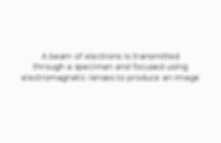
Outline how a TEM works
A beam of electrons is transmitted through a speciman and focused using electromagnetic lenses to produce an image
5 of 37
State the features of the images produced from light microscopes
In colour, low resolution, low magnifaction, 2D
6 of 37
State the features of the images produced from SEMs
No colour, good resolution, good magnifaction, 2D
7 of 37
State the features of the images produced from TEMs
No colour, best resolution, high magnifaction, 2D
8 of 37
State the features of the images produced from laser scanning confocal microscopes
Can be in colour (dyed), low resolution, low mganification, can be 2D or 3D
9 of 37
Identify the type of microscope used when presented with a photomicrograph
Laser scanning confocal microscope
10 of 37
Explain how to use a light microscope to view a specimen at low and high powers
Put the slide under the lowest power objective lens, and turn the coarse focusing knob until it's roughly in focus, then turn the fine focusing knob until you can see the specimen very clearly - switch to the higher power objective lens and repeat
11 of 37
Describe how to produce a temporary wet mount of living tissue
Put the specimen in a slide and immerse it in a drop of water, then place a cover slip on top, trying not to trap any air bubbles in
12 of 37
Describe and explain the characteristics of a good slide preparation
Specimen must be thin, slide and cover slip must be thin, no air bubbles should be trapped - all of these mean there is nothing obstructing the view of the specimen
13 of 37
Explain why slide preparations must be thin
So there aren't too many layers of cells, and the light can pass through the specimen very easily
14 of 37
Explain how to use a stage micrometer to work out the distance represented by the small divisions in an eyepiece graticule under 3 objective lenses
Divide the known length of a micrometer division by the number of small divisions on the eyepiece graticule that are within that division - repeat with different objective lenses
15 of 37
Explain how to use a stage micrometer and eye-piece to add a scale bar to a drawing
Work out the actual length of an eyepiece graticule division, then measure the width of a certain cell or tissue using this - then draw a scale bar across the cell/tissue, labelling the actual length on it
16 of 37
Explain how to use a stage micrometer amd eye-piece graticule to calculate the size of a specimen
Work out the actual length of an eye-piece graticule division, align this with the specimen, count how many divisions the specimen takes up and multiply this by the length of each division
17 of 37
Describe how to choose an appropriate number of significant figures, or decimal places to present data
Use the same number of significant figures as the divisions are
18 of 37
Explain how an adjusyment to the 'plane of focus' can alter what is viewed in a cell
Cells are in 3D, but we only see them in 2D, so if the 'plane of focus' is adjusted, the cells will be viewed from a different angle, and could be blocked by other cells, which would alter what is seen
19 of 37
Explain how a tissue slice might be misleading due to the very thin nature of the slice
It may not have all the cells present in a larger tissue slice
20 of 37
Explain why staining is useful for light microscopy
It enables us to see contrast in cells more easily
21 of 37
Describe the properties a stain needs to have to be useful for light microscopy
It should be brightly coloured, and distinctive from the rest of the cell's colour - it should also be able to stain only certain parts of a cell, not the whole thing
22 of 37
Describe how to prepare a stained specimen for viewing under a light microscope
A sample is placed on a slide and allowed to air-dry, then is heat-fixed by being passed through a flame - the specimen will adhere to the microscope slide and then take up stains
23 of 37
Name 2 common stains and the molecules they bind to
Iodine binds to starch, and crystal violet is attracted to negatively-charged material in the cytoplasm
24 of 37
State the rules for biological drawings
Include a title, state the magnification, use a sharp pencil for drawings and labels, use unlined paper, use as much of the paper as possible, draw smooth, continuous lines, don't shade, draw clearly defined structures, with correct proportions
25 of 37
State the magnification formula
Magnification = size of image / size of object
26 of 37
Explain the usefulness of a 'triangle diagram' for a simple equation'
It is very easy to visualise and work easily with what you're given
27 of 37
Explain how to calculate the magnification of an image using the magnification formular
Size of image / size of object
28 of 37
Explain how to calculate the actual size of an object using the magnification formula
Size of image / magnification
29 of 37
State the symbols used for millimetres, micrometres and nanometres
Millimetres = mm, micrometres = μm, nanometres = nm
30 of 37
Describe ways to estimate results to 'sense check' that calculated values are appropriate
Round each number in the calculation to 1 or 2 significant figures and do the calculation - the answer should be roughly the answer to the actual calculation
31 of 37
Define the term 'resolution'
The ability to see 2 objects close together as separate objects, alowing you to see the ultrastructure of cells
32 of 37
Define the term 'magnification'
The number of times that an image is larger than the object
33 of 37
State the difference between magnification and resolution
Magnification is simply how large an image is compared to the size of the object, and does not determine how much detail can be seen, whereas resolution does
34 of 37
State the resolution and useful maximum magnification of light microscopes
Resolution = 200nm, Magnigication = x 1500-2000
35 of 37
State the resolution and useful maximum magnification of SEMs
Resolution = 3-10nm, Magnification = x 100,000-500,000
36 of 37
State the resolution and useful maximum magnification of TEMs
Resolution = 0.2-0.5nm, Magnification = x 500,000-2,000,000
37 of 37
Other cards in this set
Card 2
Front
Name 4 different types of microscope
Back
Compound light microscopes, laser scanning confocal microscopes, transmission electron microscopes and scanning electron microscopes
Card 3
Front
State what SEM and TEM are abbreviations for
Back

Card 4
Front
Outline how an SEM works
Back

Card 5
Front
Outline how a TEM works
Back
Related discussions on The Student Room
- Biology microscopy »
- Memorising »
- Why is x-ray diffraction not considered a form of microscopy? BIOLOGY HELP :) »
- Career in science and research »
- exams 2022 »
- Why are some mitochondria different shapes (e.g circular) »
- GCSE Biology AQA paper 1 (May 14th) Predictions »
- Any revision tips/materials for Bio and maths? »
- Do I have to do practical endorsement for computing undergrad »
- A level Biology required practical exams »
Similar Biology resources:
5.0 / 5 based on 1 rating
Teacher recommended
0.0 / 5
0.0 / 5
0.0 / 5
3.0 / 5 based on 1 rating
5.0 / 5 based on 1 rating
2.0 / 5 based on 1 rating
4.5 / 5 based on 3 ratings
3.0 / 5 based on 1 rating
Comments
No comments have yet been made