

Biology AS1
Teacher recommended
?- Created by: Mark Kelly1
- Created on: 22-05-17 19:24
Monosaccarides
a-glucose and beta- glucose
a-glucose-hydroxyl group on bottom
beta-glucose -hydroxyl group on top
Monosaccharides have the same molecular formula but different structural formula therefore known as Structural Isomers
Disaccharides
Two monosaccharides usually Hexoses react together in a condensation reaction .This reaction is reversable and is known as Hydrolysis(adding water)
The bond formed between the two monosaccharides is known as a Glycosidic Bond

Starch Structure
Amylose= a-glucose linked by a-1,4 glycosidic bond. Coils forming a spiral held together by hydrogen bonds. Due to only a-1,4 bonds Amylose forms a long unbranched chain. Due to the presence of bulky sidegroups that cause the a-glucose molecules to lie at different angles , amylose chains form a coiled configuration
Amylopectin = a-glucose linked by a-1,4 glycosidic bonds and a-1,6 glycosidic bonds. The a-1,6 glycosidic bonds form side brances to produce a branched molecule. This branching occure as often as 1 in every 10 a-glucose monomers
Triglycerides
Combination of Glycerol and 3 Fatty Acids( Hydrocarbon Chains).
Fatty Acids are organic acids that form long hydrocarbon tailes linked to a Carboxyl Group (COOH)
Ester Bond forms between Glycerol and Hydrocarbon Tail

Saturation and Unsaturation
Saturated Fatty Acids contain the maximum number of hydrogen atoms . Linked by a C-C single bond. eg.Stearic Acid
Unsaturaturated Fatty Acids contain atleast one C=C double bonds in the chain.eg. Oleic Acid
If there is one C=C double bond it is monounsaturated.
If there is more than one C=C double bond it is Polyunsaturated
Phospholipids
One Glycerol, 2 Fatty Acids and a Phosphate.
The fatty acid molecules repel water and are insoluble in water forming hydrophobic tails whereas phosphate gives the glycerol parts of the molecule are hydrophillic properties and is soluble in Water. Phospholipids are polar molecules and this property is important in determining their orientation and function in the cell surface membrane
Proteins
Contains Carbon, Oxygen, Hydrogen,Nitrogen and Usually Sulfur
They are large Polymers formed form amino acid sub-units.
Proteins are determined by the sequence of the amino acids in the protein.
The Amino Acids differ due to them having different R-groups

Dipeptide
Amino Acids are linked together by peptide bonds . These condensation reactions with hte loss of water. The Condensation reaction takes place between the amino group of one of the amino acids and the carboxyl group of another.This link is between the Nitrogen and Carbon Atoms involved.Thebond formed is a Dipeptide.
Primary Protein Structure
The sequence of amino acids in the polypeptide chains. Its when the amino acids are linked by the peptide bonds
Secondary Protein Structure
The Orientation of how the Hydrogen bonds are orientated.
a-helix- Hydrogen bonds formed at regular intervals between amino acids. The bonds twist the chain of amino acids into a spiral or helical shape
B-pleated sheets- more rigid and less flexible configurations that the a-helix,
They are sections formed by the polypeptide chain, orientated in oppiside (anti-parrallel) directions, laying adjacent to eachother. Hydrogen bonds form between the C=O and NH groups
Nucleic Acid
Consists of
- Pentose Sugar
- Phosphate Group
- Nitrogenous Base
The three components are joined by condensation reactions./ broken down by Hydrolysis
Phosphodiester bond links pentose sugar to the phosphate (Sugar-Phosphate Backbone)
Adjacent Nucleotides can be joined together to create the Nucleic Acid. The Nucleic Acid is a chain of Nucleotides.(Polynucleotide)
DNA and RNA
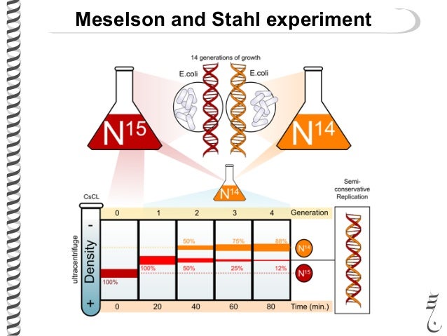
Meselson and Stahl Experiment
Meselson and Stahl tested the hypothesis of DNA replication. They cultured bacteria in a 15N medium. 15N is a heavy isotope of nitrogen so the DNA synthesized is of heavy density. They then shifted the bacteria to a 14N medium, After one generation, the DNA was all of intermediate density.
After two generations , two bands of DNA were seen, one of intermediate density and one of light density. This result is exactly what the semiconservative model predicts: half should be 15N-14N intermediate density DNA and half should be 14N-14N light density DNA. This result rules out the dispersive replication model, which predicts that after replication cycle 1, the DNA density of all DNA molecules will gradually become lower, so no intermediate density DNA should remain at 2nd Generation. The semiconservative model is correct.
Enzymes- Structure and Function
Enzymes are Biological Catalysts which speed up the rate of Metabolic Reactions.
Enzymes are globular proteins which is advantageous

Activation Energy changes
Enzymes lower the activation energy to overcome the energy barrier.
This reduction in activation energy enables reactions to take place at a more rapid rate needed to sustain life.
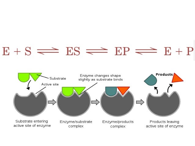
Enzyme Action
Anabolism= building up of material
Metabolism= breaking down of material
Enzymes have specific active sites ( Enzyme Specificity)
Lock and Key Hypothesis
Substrate and Enzyme Active Site complimentary.- Proves enzyme specificity
Induced Fit Model
The active site moulds itself around the substrate forming a precise fit. The active site is therefore flexible and as it changes shape to fit the substrate the enzyme is able to put pressure on the substrate breaking particular bonds and therefore lowering the activation energy required for the reaction to take place- When products released the activesite returns to pre-reaction shape.
Co-Factors
Non-protein substances that enzymes require in order to function.
Examples include Metal Ions (such as Mg2+, Ca2+ and Fe3+ ). They form attachments to the enzyme and change the shape of the active site, enabling the reaction to take place.
Co-Enzymes
Particuar Type of co-factor. They are non-protein ,organic molecules necessary ot the enzymes active. Unlike some other cofactors theyre are not permanently attached . Co-enzymes are very important in the biochemistry of respiration and photosynthesis. The coenzymes NAD and FAD act as the hydrogen acceptors in Respiration
Substrate Concentration
The Rate of reaction increases as substrate concentration increases. Substrate molecules all occupy activesites and substrate availability limits the rate of the reaction with some of the active sites unused.
The addition of extra substrate molecules has no effect as all the activesites are in use. The Number of enzyme active sites limits the rate of the reaction
Enzyme Concentration
As more enzymes and active sites become available more enzyme-substrate compleces can form and reactions can take place.
Enzyme Activity levels off as the number of substrate molecules becomming limiting.
Normally in living systems activity continues to increase as substrate is sledom limiting
Temperature
Increasing Temperature, increases the KE, molecules vibrate more vigourously and this leads to more bonds to break which effects the tertiary structure of the globular protein to be affected. This causes the Active site to change shape to cause little/no enzyme substrate complexes to form.
pH
Each enzyme has an optimum pH range in which the enzyme works best at . At either side of this optimum changes in pH will reduce the activity. This is caused by the changes in pH disrupting the bonds that are improtant in determining the protein shapr. The Ionic Bonds in particular are subject to disruption when in non opetimal pH. The further the pH is from the optimum the greater the degree of disruption to the bonding until eventuallt denaturing results.
Competitive Inhibition
Inhibitor Substances competes with the usual substrate for the active site. Competitive inhibitors are very similar in shape to the usual substrate
If the quantity of the inhibitor substance is low compared to the substrate it will have little or no effect. If it is relatively high it can significantly affect the enzyme activity
Non-Competitive Inhibititon
The inhibitor attaches itself to an allosteric site-other than the active site
The presence of the non competitive inhibitor leads to the active site changing shape so that it is no longer complementary to the substrate molecule.
Increasing the substrate concentration does not reduce the effect of the inhibitor as the two molecules are not in direct comeptition

Enzyme Immobilisation
Adsorption- Attached by weak forces onto an inert substance such as glass or a matrix.
Entrapment- trapped within polymers such as alginate beads or microspheres
Encapsulation- trapped inside a selectively permeable membrane such as Nylon
Cross-Linkage- covalenly bonded to a matrix such as Cellulose as consequnce of chemical reactions

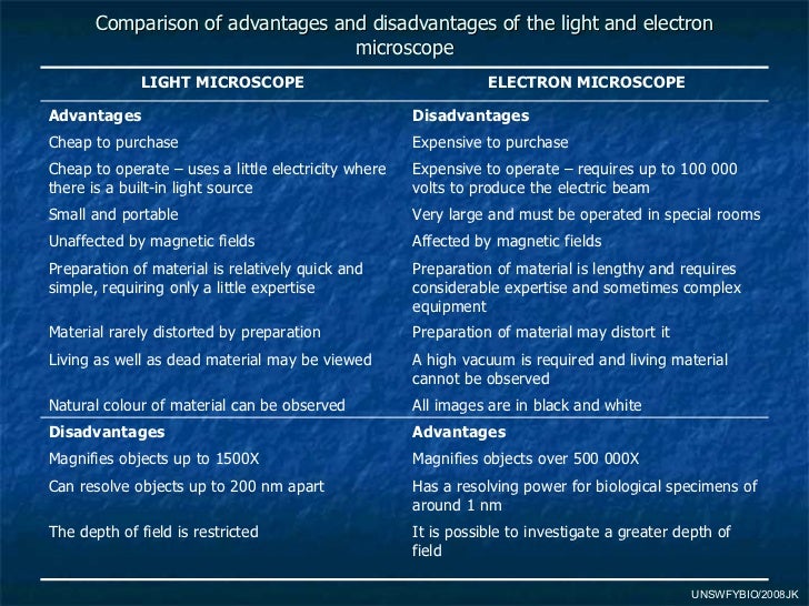
Light Microscope vs TEM
Animal Cells
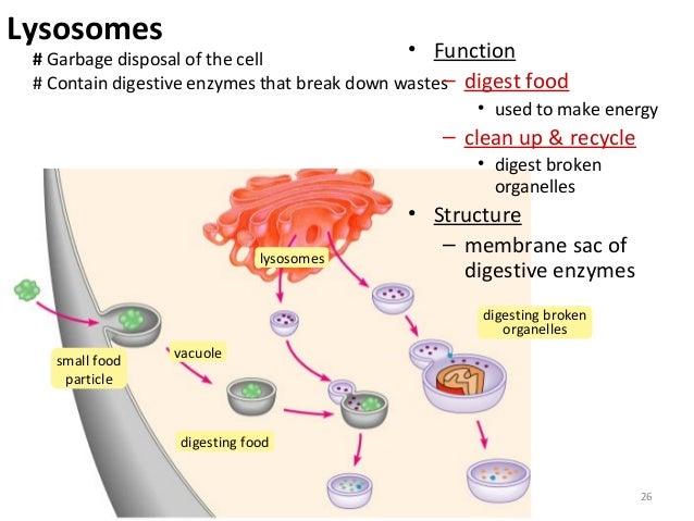
Lysosomal Activity
Mitocondria

Nucleus

Plant Cells
Plant Cell Layers

Chloroplast structure
Lipid droplets also present
Cell Surface Membrane

Function of the Phospholipid Bilayer
The backbone of the cell surface membrane
Gives the membrance its selectivity or differentially permaeble properties

Bacteriophages
Virus- commonly called phages
Normally have DNA core and are Parasitic on Bacteria
Inside their host cell the viral DNA codes for the production of new protein for new protein coats. The DNA itself replicates to make copeis that are packages within the protein coats forming new viruses .
Eventually due to the build up of viruses in the coat it ruptures releasing the viruses to continue the cell cycle

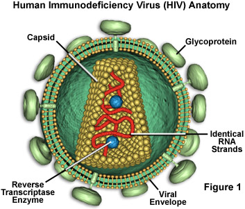
Human Immunodeficiency Virus
RNA Core.
Protein coat with a lipid bilayer containing glycoproteins.
Reverse Transcriptase enzyme catalyses the synthesis of DNA from RNA . DNA them makes new viruses by synthesising new protein coats and viral RNA
These viruses are known as Retroviruses as the viral RNA is used as a template to make DNA . This is the revers of the normal transcription process where DNA is used to make mRNA as part of the protien synthesis.
AIDS
In Humans HIV invades the lymphocyte - helper-T-cells.
These T cells are very important for the Immune System when protecting against disease.
As progressively more T-Cells are destroyed the immune system becomes critically comprimised and medical condition AIDS can develop
Diffusion
The net movement of a substance from where it is in highter concentration ot where it is in a lower concentration . Its not restricted to occuring across membranes
Factors Affecting Diffusion-
- Concentration Gradient - greater the conc. gradient faster diffusion
- Size of molecule- smaller molecule faster diffusion
- Temperature- high temp. inc diffusion
- Thickness of Exchange Surface- thin surface faster diffusion
- Surface Area of Membrane - greater SA faster diffusion
Facilitated Diffusion
Channel Proteins- Static- gated/non-gated. Allow charged particals to pass through.
Carrier Proteins- Take in the diffusing moleule such as glucose change shape and release the molecule on the other side of the membrane. These protein carriers have binding sites that match specific moleucles and they assist the movement of these molecules across the membrane
Active Transport
Moving molecules against their concentration gradient . Energy required- ATP
Active Transport involves protein carrier molecules(pumps). The substance binds to the carrier protein.
Cells that carryout Active Transport have high number of Mitocondria- important for synthesis of ATP energy

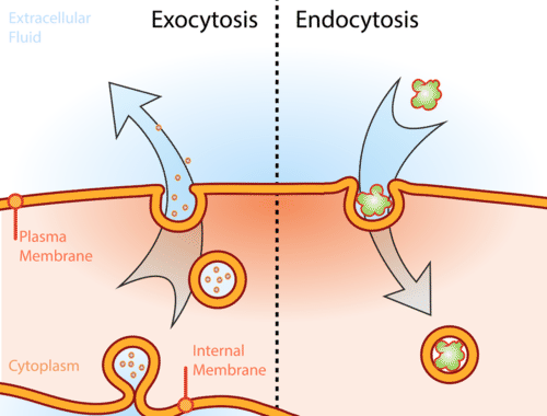
Cytosis
Endocytosis- movement of substances into the cell- cell wall invaginates and pinches off as vesicle
Exocytosis- movement of substances out of the cell-vesicle fuses with cell membrane
Phagocytosis- transporting solid material into cell
Pinocytosis- transport of fluid into cell
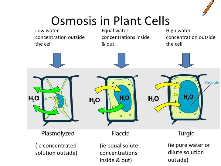
Osmosis
High water potential to lower water potential
Water potential is the tendancy to take in water by osmosis from pure water across a selectively permaeble membrane. Pure water was solute potential of 0kPa, Solutes decrease solute potential

Solute Potential is the potential of a solution to take in water Pressure Potential is the effect of pressure on the solution ( ONLY IN PLANT CELLS)
Water Potential Graphs
As cell begins to become more turgid the membrane begins to exert a force on the cell wall( Incipient Plasmolysis)At full Turgor the maximum force betweeen the cell and the cell walWater Potential of the cell is zero so no more water can enter Solute potential is still negative at full turgor as solutes are present in the cell As the pressure potential becomes positive it begins to hinder the water entering the cell therefore the cell potential and solute potential diverge As the cell takes in water by osmosis its contents become less concentrated therefore the solute potential increcreases. There is no pressure potential to restrict the water intakes therefore the solute potential=cell potential
Osmosis in Plant Cells
Osmosis in Animal Cells
Cell Cycle
Checkpoints

G0 Phase

Graph for Amount of DNA in Cell Cycle
DNA doubles during DNA replication and chromatid formation.
DNA is then Halved during Cytokinesase as DNA is divided between two daughter cells
Chromosomes
Chromasomes consist of an ectended DNA molecule sipported by special proteins called Histones. Chromatin condenses to form Visible Chromosomes during Nuclear Division
DNA coils tightly around the stack to form a structure called a Nucleosome.

Mitosis
Meiosis
Difference in Mitosis and Meiosis
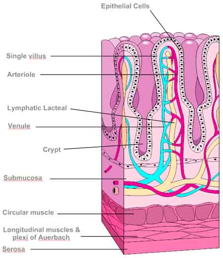
Ileum
Villus
Structure of Plant Cell
- Waxy Cuticle - Prevents water loss amd provides waterproofing
- Palisade Mesophyll Layer- photosynthesis . Rich in Chloroplasts
- Spongy Mesophyll Layer-gas exchange- intercellular spaces
- Xylem and Phloem - transport of ions and water moleucles
- Stomata - Allow gas exchange . Allows water vapour to diffuse out of the leaf
Comments
Report