GI Tract Theme 2
0.0 / 5
- Created by: Splodge97
- Created on: 22-05-17 13:49
What are unicellular glands?
Individual cells with an epithelium such as type I pneumocytes or goblet cells
1 of 81
What are the types of multicellular glands?
Comprised of different cell types - may be tubular (exocrine cells line ducts), acinar (exocrine cells in a sac at the end of a duct) or compound tubulo-acinar (exocrine cells along tubes and in a sac at the end)
2 of 81
What are the classifications of tubular glands?
Simple = exocrine cells lining one straight duct (as in Brunner's glands). Compound = exocrine cells lining branching tubes (as in gastric glands).
3 of 81
What are the classifications of acinar glands?
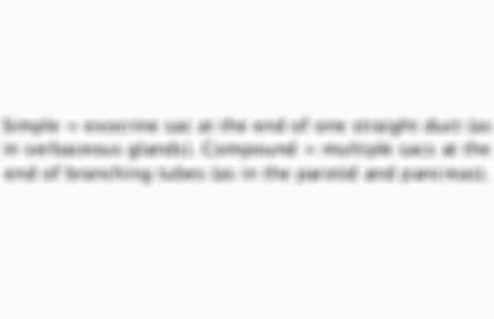
Simple = exocrine sac at the end of one straight duct (as in serbaceous glands). Compound = multiple sacs at the end of branching tubes (as in the parotid and pancreas).
4 of 81
How do different gland types secrete?
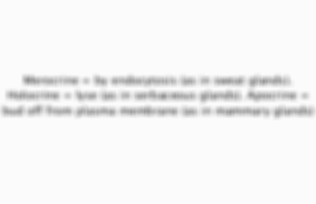
Merocrine = by endocytosis (as in sweat glands). Holocrine = lyse (as in serbaceous glands). Apocrine = bud off from plasma membrane (as in mammary glands)
5 of 81
How much saliva do we secrete each day? How can saliva flow rates be defined?
650ml. Rates = normal 27ml/h, hyposalivation = 1-4ml/h, xerostomia =
6 of 81
What are secretory end pieces?
Bunches of acini sacs present at the ends of ducts; may be serous acini or mucous alveoli
7 of 81
Describe serous acini
Spherical shape with a smaller lumen, intercalated cnanliculi which branch from intercalated ducts between their acinar cells. Apppear dark purple under H+E.
8 of 81
Describe mucous alveoli
Tubular shape, large lumen. Compressed basal nuclei in smaller cells. Appear light pink under H+E, light blue with alcian blue. Compact nuclei at edges differentiate from fat cells.
9 of 81
What are the features of interlobular excretory ducts?
Largest diameter, pseudostratified columnar epithelium (stratified cuboidal in sublingual). Small clear goblet cells within them. May contain congealed saliva/mucous (dark line) or light pink loose connective tissue.
10 of 81
What are the features of intralobular striated ducts?
Intermediate diameter, lined with simple columnar epithelium (simple cuboidal in sublingual). Striped as folded membrane to aid reabsorption/secretion of electrolytes (HCO3-).
11 of 81
What are the features of intralobular intercalated ducts?
Lumen continuous with that of the secretory end piece. Lined with simple cuboidal epithelium, longer in the parotid and shorter in the submandibular. Provide lysozymes and lactoferrin to saliva.
12 of 81
What are myoepithelial cells?
Star shaped contractile cells joined by desmosomes, contract to expel secretions from secretory end pieces by exocytosis. Maintain polarity/structural integrity of acini and have tumour supressor activity.
13 of 81
What are serous demilunes?
Artefacts in mixed mucous glands (mainly submandibular). Tissue fixation causes swelling of the serous cells so they are compressed into cresents at the extremes of secretory end pieces. Blue in middle with purple serous cells around the edges.
14 of 81
How can H+E staining determine the stage of the cell cycle?
Cells in the early stage have darker cytoplasm (as rich in RNA/ribosomes); becomes pinker towards end of cycle
15 of 81
Describe the histology of the parotid gland
Largest, compound acinar, mainly serous (so many serous acini/striated ducts). Stensen's duct. Lobular arrangement with fibrous septum (containing excretory ducts/blood vessels) between them. Few/no fat cells, mucous alveoli or serous demilunes.
16 of 81
Describe the histology of the submandibular gland
Mixed, compound tubulo-acinar, mainly serous. Wharton's duct. Contains serous acini and mucous alveoli, fact cells and serous demilunes. Lobules not as clearly separated.
17 of 81
Describe the histology of the sublingual gland
Smallest, compound tubulo-acinar, mixed (mainly mucous). Ducts of Rivinus. Large excretory (stratified cuboidal), smaller intralobular so can't differentiate (simple cuboidal like canaliculi). Many mucous alveoli, no serous demilunes/acini.
18 of 81
What are the features of the minor salivary glands?
600-1000, present as aggregates in submusous layer. Mixed but mainly mucous (except Von Ebner's glands, mainly serous). May be labial, in the cheeks or palatal (comprise most of soft palate).
19 of 81
What is the function of saliva?
Forms food bolus by moistening and lubricating the oral cavity
20 of 81
What stimulates saliva secretion?
Parasympathetic stimulates voluminous saliva (serous, from parotid, collected using Lashley's canula/Curby cup), sympathetic releases viscous saliva (more mucous, from sublingual/submandibular, collected using Schneyer's apparatus)
21 of 81
What are the organic components of saliva?
Proteins (A-amylase, mucins, antimicrobial proteins, immunoglobulin A (prevents viral/fungal infection)), glycoproteins, glycosaminoglycans, carbohydrates and lipids
22 of 81
What anti-microbial proteins are present in saliva?
Lysozymes (lyse bacteria) and lactoferrin (bind iron needed for bacterial cell growth)
23 of 81
What are the inorganic components of saliva?
H+ (alters pH, shift equilibrium for calcium phosphate), Ca2+ (actively transported), Pi (alters solubility of calcium phosphate, buffers) and fluoride (undergoes facilitated diffusion, speeds enamel remineralisation)
24 of 81
What stimulates saliva production?
Increases upon aromas, mechanical/chemical stimulation. Secretions from submandibular (from 0.26ml/min) and parotid (from 0.12 ml/min) triple; only double from sublingual/minor (from 0.12ml/min)
25 of 81
What are preblems associated with xerostomia?
Difficulty eating/swallowing, regular drinking, hoarseness, food between teeth, no protection from acidic foods (may cause oral thrush/gingival recession/tooth loss)
26 of 81
What are the causes of xerostomia?
Medications (CNS depressants, CVS drugs, muscle relaxants), diseases (Sjogren syndrome, Parkinsons, cyctic fibrosis, uncontrolled diabetes) or radiation/chemotherapy, bulimia, depression, salivary stones and ageing.
27 of 81
What are the functions of the stomach?
Store chyme (4-5 hrs) to slow passage into SI (5-10ml past pyloric sphincter increasing absorption). Ancrum digests (chemical via H+, pepsin and gastric lipase, mechanical through churning every 15-20sec). pH 1-2 protective (lyses bacteria).
28 of 81
How does H+ perform digestion?
Unfolds globular proteins into polypeptide chains, activates pepsinogen to pepsin
29 of 81
Does the stomach function in absorption?
Yes, a little - absorbs cold water, aspirin, electrolytes and alcohol (alcohol dehydrogenase in gastric mucosal cells converts to acetaldehyde)
30 of 81
How is auto-digestion in the stomach prevented?
Release of HCO3- from gastric glands to form a protective layer around the epithelium (increased in inflammation/presence of irritants like alcohol)
31 of 81
How do parietal cells secrete gastric acid?
Secrete H+ via active tranport against strong concentration gradient (as pH in ICF is 7.2); Cl- follows through open channels along its electrochemical gradient
32 of 81
What drugs are given to treat acid reflux?
H2 receptor antagonists (compete with histamine, stain tongue/cause sores), proton pump inhibitors (block AT of H+, cause dry mouth), prostaglandin analogues and antacids (cause HCO3-/mucous, chalky taste), chelates (coat mucosa, cause dry mouth)
33 of 81
What are long and short reflexes?
Long = coordinated by CNS, stimulated by sensory information before food is digested (indirectly activating GI tract via ANS); activated by changes in digestion so ANS can modify. Short = ENS coordinates, stretch/chemo receptors of GI tract activate
34 of 81
What occurs in the cephalic phase of digestion?
Sensory stimui created by the presence of foood activate long reflexes, cause parasympathetic (via vagus) to increase stomach muscle/gland activity (bad taste/smell does opposite via sympathetic)
35 of 81
What occurs in the gastric phase of digestion?
Stretch/chemoreceptors detect food (caffiene/protein stimulating most). G cells secrete gastrin to blood (increases chuurning , gland secretion and sphincter relaxation). H+ increase activates chief cells to secrete pepsinogen (converted to pepsin).
36 of 81
How does gastrin increase glandular secretion?
Directly stimulates parietal cells and also activates ECL (enterocromaffin like) cells to secrete histamine (acts upon parietal cells)
37 of 81
What negative feedback occurs after the gastric phase?
H+ stimulates D cells to release somatostatin; inactivates G cells, chief calls and parietal cells (so pH rises/digestion stops)
38 of 81
What occurs in the intestinal phase of digestion?
Release of chyme into duodenum, stretch/chemo receptors sense distension, fatty acids and sugar so slow stomach/increase intestinal activity via sympathetic ANS and short reflexes. H+ stimulates secretin, fats/proteins stimulate CCK, carbs GIP.
39 of 81
What is the action of secretin in the intestinal phase?
Released from enteroendocrine cells of SI; it reduces gastric secretions/motility and HCO3- release for neutralisation (removing the stimulant for its release)
40 of 81
What is the action of cholesystokinin in the intestinal phase?
Released from enteroendocrine cells of the SI; inhibits acid secretion/gastric motility and causes pancreatic acinar cells to secrete preoteases and lipases
41 of 81
What is the action of gastric inhibitory peptide in the intestinal phase?
Released from enteroendocrine cells of the SI; decreases acid secretions and stimulates insulin to deal with glucose increase
42 of 81
What are the functions of the small intestine?
Weak peristalsis so chyme stays for 3-5hrs, maximising absorption. Moves food along GI tract by increasing activity more with distension. Digests proteins (as pepsin partially hydrolyses), lipids and starch. Neutralises, produces mucous and absorbs.
43 of 81
How is intestinal motility decreased upon defaecation?
Via the sympathetic ANS
44 of 81
How does the SI perform neutralisation of gastric acid?
Via bile secreted into the duodenum
45 of 81
How is mucous generated in the SI?
From goblet cells which release it for protection/lubrication when stimulated by vasoactive intestinal polypeptide
46 of 81
How is the SI adapted for absorption?
Tight junctions between its columnar epithelia (made of claudin and occludin proteins) as well as adherens junctions and desmosomes; protective function as pathogens can't enter (must cross apical/basolateral membranes by AT/facilitated diffusion)
47 of 81
What is the action of sucrase isomaltase at the brush border membrane?
Hydrolyses sucrose to fructose and maltose and converts aplha limit dextrins to single AA's
48 of 81
What is the action of lactase at the brush border membrane?
Hydrolyses lactose to glucose and galactose - expression reduces upon weaning (deficiency causing lactose intolerance)
49 of 81
What is the action of peptidases at the brush border membrane?
These act on a mixture of small peptides to produce a mixture of AA's and di/tri peptides
50 of 81
What is the action of enterokinase at the brush border membrane?
Partially hydrolyses zymogen proteases to their active form - notably hydrolyses trypsinogen to trypsin (which activates a large number of pancreatic enzymes)
51 of 81
What is the function of the square acinar cells of the pancreas?
Produce zymogens of proteases, amylases, lipases and nucleases which are released when CCK binds to them at the basolateral membrane
52 of 81
What is the function of the square centro-acinar cells of the pancreas?
Modify the ionic composition of pancreatic secretions (releasing HC03- for neutralisation via carbonic anhydrase). Stimulated to release their contents by binding of secretin at their basolateral membrane
53 of 81
What active enzymes does the pancreas secrete?
Phospholipase A3 (digests phospholipids), amylases (yield maltose, maltotriose and A-limit dextrins) lipases (hydrolyse triglycerides to 2-monoglyceride and 2NEFA's), ribonuclease, deoxyribonuclease, gelatinase and elastase
54 of 81
What are basket cells?
Surround the acinar cells of the pancreas and contract to intiate secretion by exocytosis (as in myoepithelial cells of slaivary glands)
55 of 81
How does the ANS stimulate pancreatic secretions?
Vagus nerve (as part of parasympathetic ANS) increases secretion in anticipation of a meal
56 of 81
What stimulates the secretion of bile?
Secretin (directly) and CCK (indirectly by causing relaxation of sphincter of Oddi and contraction of gall bladder)
57 of 81
What does bile contain?
Conjugated bilirubin, bile acids (cholesterol derivatives), bile salts (aid fat digestion by forming micelles, are recycled), glucuronide, glutathione conjugates, lipids and enzymes from hepatocytes
58 of 81
What are the functions of the large intestine?
Mechanical digestion (greater peristaltic waves as thicker muscular externa, 3-12/min). Bacteria within it ferment chyme (makes CO2 + CH4 from carbs, odorous indoles from proteins and brown pigments from bilirubin). Removes 90% of water in 3-10hrs.
59 of 81
What is haustral churning?
Occurs in the large intestine - pouches are filled from below by elevator contractions
60 of 81
What is the gastroilial reflex
Causes release of gastrin when stomach full to relax iliocaecal sphincter (so SI empties into LI) - same time as gastrocolic reflex
61 of 81
What is the gastrocolic reflex? What occurs after it?
A strong peristaltic wave, moves contents of transverse colon into rectum. Defaecation then occurs when parasympathetic ANS causes rectum to contract and relaxation of internal sphincter.
62 of 81
How does pancreatic insufficiency increase the risk of excessive bleeding?
Lack of pancreatic lipase means chylomicrons not formed, prevents digestion of vitamin K for use in blood clotting
63 of 81
What causes pancreatic insufficiency?
Viruses like HIV, alcohol and gall stones
64 of 81
Describe the arrangement of hepatocytes in the liver
Radiate as a plate from a central vein so they can contact the bloodstream on one side and bile canaliculi on the other. Bile canaliculi project from the main bile duct and so collect bile from hepatocytes and transport it to the gall bladder.
65 of 81
What are sinusoids?
Vascular spaces between hepatocytes (open spaces forming sinusoid capillaries that blood can run through)
66 of 81
What are kupffer cells?
Between hepatocytes, act as phagocytes to kill bacteria that have entered the blood from the GI tract
67 of 81
What is a portal triad?
Section of the liver where the hepatic artery, hepatic vein and bile duct run alongide each other
68 of 81
How does the liver function in detoxification?
Removes exogenous toxins/endogenous substances at high levels; either excretes in bile, kupffer cells phagocytose or hepatocytes modify (as in conversion of glutamate to A-ketoglutarate to form NH4+ for the urea cycle)
69 of 81
How does the liver function in protein synthesis?
Produces albumin (carrier, regulates oncotic pressure), golbulin (transports cholesterol/hormones, produces clotting factors), fibrinogen (for clotting), transferrin (for Fe transport) and lipoprotiens (transport triglycerides/cholesterol)
70 of 81
How does the liver function in carbohydrate metabolism?
Site of glycogenesis, glycogenolysis (both liver and muscles) and gluconeogensis (just liver)
71 of 81
How does the liver function in lipid metabolism?
Chylomicrons transport fats to the tissues, forming HDL cholesterol and chylomicron remnants when triggered by lipoprotein lipase. In liver used to form cholesterol, NEFA's and other lipoproteins (form LDL cholesterol). Cholesterol used to make bile.
72 of 81
What drugs does the liver process?
Miconazole (anti-fungal to treat oral thrush), antibiotics like tetracycline, paracetamol, NSAID's and metronidazole (also anti-fungal)
73 of 81
What is bilirubin and how is it secreted?
Made in liver, bone marrow and spleen from haem groups (without Fe). Combines with glucuronic acid to form conjugated bilirubin. Secreted into bile and converted to urobilinogen by bacteria of LI; absorbed and recycled or excreted in urine.
74 of 81
How does vomiting act as an immune function of the gut?
Reflex controlled by emetic centre of the brain, initiated by abnormal food in stomach. Parasymathetic ANS and ENS cause increased saliva and retroperistalsis. Lower intrathoracic pressure/increase in abdominal pressure propel stomach contents.
75 of 81
What sympathtic responses occur during vomiting?
Inrease in HR and sweating
76 of 81
How does GALT (gut-associated lymphoid tissue) act as part of the immune function of the gut?
Acts if pathogens cross mucosal-epithelial barrier. Present as lymph nodes in the tonsils and appendix and Peyer's patches in the SI at the start of the submucosa.
77 of 81
What do Peyer's patches contain? How do they act?
Contain macrophages, dendritic cells and lymphocytes. M cells of SI (with clathrin-coated pits containing receptors attracting bacteria) signal Peyer's patches upon pathogens binding so after transcytosis of the pathogen GALT is activated.
78 of 81
What occurs when GALT is activated once Peyer's patches have been signalled?
Interstitial fluid macrophages, lymphocytes and cytokines are released to trigger an inflammatory response and increase Cl-, fluid and mucous to destroy the pathogens. Immunoglobulins also secreted into the intestine to destroy remaining bacteria.
79 of 81
How do Salmonella and Shigella cause food poisoning?
Have adapted receptors which aren't recognised by M cells as pathogenic so can perform transcytosis without causing an immune response (causing diarrhoea/vomiting in an effort to remove from the GI tract)
80 of 81
How do LI bacteria act as our final immune defence?
During their competition with foreign bacteria they release antimicrobial proteins like lysozymes (destroy gram positive bacteria), lactoferrin and defensins (anti-viral). It is thought these could be harnessed to replace antibiotics.
81 of 81
Other cards in this set
Card 2
Front
What are the types of multicellular glands?
Back
Comprised of different cell types - may be tubular (exocrine cells line ducts), acinar (exocrine cells in a sac at the end of a duct) or compound tubulo-acinar (exocrine cells along tubes and in a sac at the end)
Card 3
Front
What are the classifications of tubular glands?
Back

Card 4
Front
What are the classifications of acinar glands?
Back
Card 5
Front
How do different gland types secrete?
Back
Related discussions on The Student Room
- A&P revision recommendations »
- Edexcel A Level History Paper 1 (9HI0 1A-1H) - 24th May 2023 [Exam Chat] »
- Just a random medic blog tbh ;-; »
- A-level geography NEA »
- The movement of substances within living organisms. »
- Jekyll & Hyde Predictions »
- dissertation topic »
- 1984 and the Handmaid’s tale »
- Books to compare (for coursework/comparative essays) »
- Gcse English lit poetry »
Similar Dentistry resources:
0.0 / 5
0.0 / 5
0.0 / 5
0.0 / 5
0.0 / 5
0.0 / 5
0.0 / 5
0.0 / 5
Comments
No comments have yet been made