CVS Theme 2
0.0 / 5
- Created by: Splodge97
- Created on: 20-05-17 16:17
What is the myocardium?
The thick layer of cardiac muscle cells (cardiomyocytes) beneath the visceral pericardium. It contracts/relaxes and maintains the shape of the heart chambers.
1 of 71
What is the endocardium?
Thin layer of endocardial cells constituting the blood-tissue barrier of the heart; it secretes signalling molecules which control cardiac function (e.g. the ion concentration surrounding cardiomyocytes)
2 of 71
What are the features of cardiomyocytes?
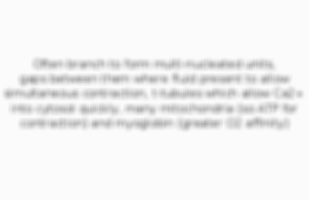
Often branch to form multi-nucleated units, gaps between them where fluid present to allow simultaneous contraction, t-tubules which allow Ca2+ into cytosol quickly, many mitochondria (so ATP for contraction) and myoglobin (greater O2 affinity)
3 of 71
What are the features of intercalated discs?
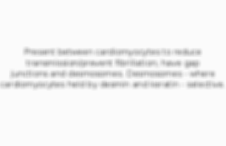
Present between cardiomyocytes to reduce transmission/prevent fibrillation; have gap junctions and desmosomes. Desmosomes - where cardiomyocytes held by desmin and keratin - selective.
4 of 71
Why do autorhymic cells set different beat frequencies? What are the different types of autorythmic cells?
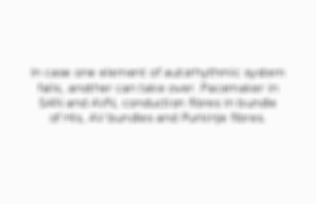
In case one element of autorhythmic system fails, another can take over. Pacemaker in SAN and AVN, conduction fibres in bundle of His, AV bundles and Purkinje fibres.
5 of 71
What rate does the sino-atrial node set?
70-100bpm (though normally lower through continuous discharge of parasymppathetic axons from vagus). Strong sympathetic innervation increases to 80-120bpm, strong vagal reduces to 20-40bpm (in sleep as 70% blood in veins/contractility reduced 20-30%)
6 of 71
What heart rate is set by the atrio-ventricular node?
40-60bpm; also delays the impulse to allow atria time to fully fill
7 of 71
What heart rate is set by the Bundle of His and AV bundles?
20-40bpm; note impulse only travels at 1m/s in AV bundles so ventricles can fully fill
8 of 71
What heart rate is set by the Purkinje fibres?
15-30bpm, though conductance velocity is 5m/s (so rapid ventricular contraction)
9 of 71
What occurs in cardiomyocyte contraction?
Threshold met by Na+ influx --> Na+ VGC's open, depolarisation --> Ca2+ VGC's triggered to open (by binding of DHP) --> calcium induced calcium release from SR (binds to ryanodine receptors) --> 300nM Ca2+ initiates contractile apparatus
10 of 71
What occurs in cardiomyocyte relaxation?
Ca2+ actively back into SR by SERCA and ECF by membrane Ca2+ATPase. NCX at cell membrane actively removes 3Na+ and intakes Ca2+, Na+/K+/ATPase removes 3Na+ and intakes 2K+; action of these electrogenic as NCX works faster so positive influx overall
11 of 71
Where does ATP bind to SERCA?
Specific nucleotide binding domain (causing shape change)
12 of 71
What normally inhibits SERCA?
Phospholamban (PLB) attached to it on the SR membrane; phosphorylated by PKA after cAMP pathway activated (activating SERCA)
13 of 71
What is pulse pressure?
Difference between diastolic pressure (80mmHg, for 2/3 of cardiac cycle) and systolic pressure (120mmHg, for 1/3 of cardiac cycle); as such normally 40mmHg
14 of 71
How is mean arterial pressure calculated?
Diastolic pressure + 1/3 of pulse pressure (usually 93.3mmHg)
15 of 71
What occurs during the atrial contraction stage (of active systole)?
Blood moves from atria into ventricles via AV valves
16 of 71
What occurs during the isovolumetric contraction stage (of active systole)?
First contraction of the ventricles, increasing pressure in them to close the AV valves
17 of 71
What occurs during the ventricular ejection stage (of active systole)?
Ventricles undergo their final contraction, raising pressure in them to open the SL valves
18 of 71
What occurs during the isovolumetric relaxation stage (of passive diastole)?
All chambers of heart relaxed, decreasing pressure in ventricles so SL valves close. Some blood still in ventricles after ventricular ejection.
19 of 71
What occurs during the atrial filling stage (of passive diastole)?
Atria fill, some blood leaking through AV valves into ventricles
20 of 71
What occurs during the ventricular filling stage (of passive diastole)?
AV valves open due to increased pressure in the atria upon filling, so blood moves into the ventricles. All chambers contain blood.
21 of 71
What is cardiac output?
Amount of blood pumped out of the heart per minute, usually 5L/min (2Ol/min in exercise, 35L/min in athletes through hypertrophy). Calculated by CO = HR x SV (blood pumped out per cardiac cycle in ml, so SV=EDV-ESV). CO should equal venous return.
22 of 71
What is end diastolic volume (EDV)?
Volume of blood in the ventricles before systole (when they are full)
23 of 71
What is end systolic volume (ESV)?
Volume of blood in ventricles after systole (when at its lowest). Can diagnose aortic insufficiency (regurgitation of blood into ventricles from SL valves).
24 of 71
What is the ejection fraction?
Proportion of blood ejected from a ventricle per peartbeat; calculated by EF=SV/EDV
25 of 71
How does Starling's law explain why venous return and cardiac output are equal?
The ventricluar walls stretch in proportion to the venous return, so the elastic energy within them to initiate contraction generates an equal cardiac output (similar to arterial recoil in haemodynamics/study of blood flow)
26 of 71
How does acetylcholine affect the heart?
Released from the vagus nerve to the SAN, increases K+ permeability by opening K+ channels to allow efflux; as such cardiomyocytes become hyperpolarised so greater stimulus needed to initiate contraction (decreasing bpm/increasing repolarisation)
27 of 71
How does nor-adrenaline affect the heart?
Released from the sympathetic chain to the SAN, increases Na+ and Ca2+ permeability (opening channels to allow influx) so cardiomyoocytes depolarised, reducing the stimulus needed for contraction (increasing bpm/reducing repolarisation)
28 of 71
What is electrophysiology?
Study of the electrical properties of biological cells, tissues and organs. Electrophysiological recordings (extracellular and intracellular) should be taken in a Faraday cage.
29 of 71
What can extracellular recordings obtain?
Frequency of action potenitals across multiple cells (multi-unit) or a single cell (single-unit)
30 of 71
What can intracellular recordings obtain?
Membrane potential, using current-clamp (current controlled so voltage measured) or voltage clamp (voltage controlled so current measured) techniques
31 of 71
How are sharp electrode impalements and the perforated patch technique used to gain intracellular recordings?
Sharp impalements more invasive so cell can't function after, perforated patch less invasive (firm contacts without impalement) so cell can function after and electrode much larger so can gain whole-cell values
32 of 71
What is membrane capacitance?
Ability of the membrane to store electric charge which is present as the electrochemical gradient created by ions (making it passive). It is measured as the resistance to an ion's electrochemical gradient within its ion channel.
33 of 71
What is membrane conductance (G)?
This is the degree to which the membrane conducts electricity, which is the reciprocal of resistance (so G=I/V)
34 of 71
Describe the apperance of graphs for the transmembrane current of K+
Produce a wide outward current with a decreasing gradient (as K+ flow activated slowly and inhibited more slowly)
35 of 71
Describe the apperance of graphs for the transmembrane current of Na+
Produce a narrow inward current (with a sharp, rapid decrease and increase) as Na+ flow is both activated and inactivated quickly
36 of 71
What occurs in phase 0 of an action potential?
First stage (depolarisation) - threshold is reached, Na+ VGC's open, depolarisation, more Na+ VGC's open, peak at +40mV
37 of 71
What occurs in phase 1 of an action potential?
Early repolarisation - Na+ VGC's close rapidly, K+ VGC's open slowly. Represented as small negative gradient after peak.
38 of 71
What occurs in phase 2 of an action potential?
Plateau phase - Ca2+ release from the SR to initiate contraction nullifies the effect of K+ efflux via K+ VGC's
39 of 71
What occurs in phase 3 of an action potential?
Rapid repolarisation - Ca2+ removed to ECF/back into SR so K+ efflux again dominates, causing repolarisation then hyperpolarisation (as K+ VGC's too slow to close)
40 of 71
What occurs in phase 4 of an action potential?
Resting potential - K+ leakage channels dominate, polarisation/baseline maintained by Na+/K+/ATPase
41 of 71
Describe the appearance of AP's in the pacemakers (SAN and AVN)
Start with a slope, then sharp rise followed by gradual repolarisation. No plateau phase.
42 of 71
Describe the appearance of AP's in the atria
Similar to the pacemakers but baseline at start and minimal plateau phase. Walls thin so short AP's/lesser Na+ rise for contraction. Minimal plateau as have type T (open at generator potential) and type L (open at AP) Ca2+ channels so rapid removal.
43 of 71
Describe the apperance of AP's in the bundle of His and AV bundles
Start from baseline with longer period before depolarisation (due to fibre composition). Action potentials are longer, with a plateau phase.
44 of 71
Describe the apperance of AP's in the Purkinje fibres and ventricles
Occur slowly and last longer (since longer Ca2+ influx to contract thicker walls), peak higher (greater Na+ influx) starting from baseline with a long plateau phase (as only type L Ca2+ channels so slow removal - though aids greater contraction)
45 of 71
How is pace-making current (determines the heart rate) generated?
Influx of nor-adrenaline into the SAN activates adenylate cyclase, cAMP generated; binds to HCN (hyperpolarisation cyclic nucleotide channel) which allows an Na+ influx (generator potential). Acetylcholine inactivates adenylate cyclase, slowing bpm.
46 of 71
What is an ionotropic effect?
A change in Ca2+ concentration and contractility. Parasympathomimetics = negatively ionotropic (as decrease contractile force), sympathomimetics = positively ionotropic (as increase contractile force).
47 of 71
What is a chronotropic effect?
Change in heart rate. Parasympathomimetics = negatively chronotropic (as decrease bpm), sympathomimetics = positively chronotropic (as increase bpm).
48 of 71
What is a domotropic effect?
Change in contraction speed/rate of contractile pulses. Parasympathomimetics = negatively domotropic (as decrease contraction speed/pulse rate), sympathomimetics = positively domotropic (as increase contraction speed/pulse rate).
49 of 71
Describe how an ECG is recorded
Requires 12 leads - three limb leads (I, II and III), three augmented leads (use Goldman's central terminal as their negative electrode), six pericardial leads (measure perpendicular plane) and rhythm ***** (one lead's ECG so many can be analysed)
50 of 71
What is a positive deflection?
A peak from isoelectric baseline in an ECG caused by depolarisation towards the positive electrode or repolarisation away from the positive electrode
51 of 71
What is a negative deflection?
A dip from isoelectric baseline in an ECG caused by repolarisation towards the positive electrode or depolarisation away from the positive electrode
52 of 71
What are the main features of an ECG?
P wave (atrial depolarisation), QRS complex (ventricular depolarisation), T wave (ventricular repolarisation) and U wave (repolarisation of papillary muscle and Purkinje fibres)
53 of 71
What intervals occur in an ECG?
PR interval (between start of P wave and start of QRS complex) and QT interval (between start of QRS and end of T wave)
54 of 71
What segments occur in an ECG?
PR segment (level period between end of P wave and start of QRS, where atria contract) and ST segment (level period between end of S phase and start of T wave, where ventricles contract)
55 of 71
How is heart rate calculated using papaer parameters?
Time the distance between the P waves equates to gives artial contraction rate, time the distance between the R peaks represents gives the ventricular contraction rate. Divide these times by 60 seconds.
56 of 71
What is a standard calibration marker?
A rectangular peak indicating the ECG must calibrate/is calibrated incorrectly
57 of 71
What is somatic (mesodermal) muscle interferance?
A slightly flucuating isoelectric baseline (with peaks still clear) due to shivering/nervousness
58 of 71
What occurs when an ECG is taken during muscle tremors (caused by neuromuscular disease)?
Wide, large fluctuations in the isoelectric baseline where P,T and U waves can't be distinguished
59 of 71
What is the cause of a sloping baseline?
The patient is standing (they should be horizontal and relaxed)
60 of 71
What is a patient movement artefact?
Where ECG peaks are undefined, making heart rate difficult to calculate, due to excessive movement
61 of 71
What does mains interferance cause in an ECG?
A thick 'noisy' isoelectric baseline, should be moved/put in a Faraday cage
62 of 71
What is sinus arrythmia?
Normal increase in heart rate upon inspiration (decreasing distance between P waves/R peaks) and decrease upon expiration (increasing the distance between P waves/R peaks) - it is more profound in children
63 of 71
What is sinus bradycardia (bradyarrythmia)?
Slow heart rate (below 50bpm) which may cause fatigue/weakness/dizziness. It is characterised by long baseline periods between ECG sequences.
64 of 71
What is sinus tachycardia (tachyarrythmia)?
This is a heart rate above 100bpm which is normal in exercise but abnormal in cardiac pathology. It is characterised by short/no baseline periods between ECG's.
65 of 71
What is atrial fibrillation?
Rapid/irregular beating which starts as short periods that become longer, possibly constant. Lack of clear P waves (as atrial activation 300-600bpm) and longer periods between R peaks (most impulses meet AVN in refractory period).
66 of 71
What is ventricular fibrillation?
Where impulses are rapidly generated in one/more parts of the ventricles so venticular muscle activity and heart rate is uncoordinated (making very serious). P waves unrecognisable, P-R intervals/QRS complexes absent and R peaks close (HR >300bpm).
67 of 71
What occurs in a premature atrial beat?
A heartbeat of the atria occurs early, resulting in an early, abnormal P wave
68 of 71
What occurs in a premature ventricular beat?
Here an abnormal impulse occurs from within the ventricles resulting in an early R peak followed by a pause. QRS complex is often wide and abnormal, with the T wave occuring in the opposite direction to it.
69 of 71
What occurs in ventricular tachycardia?
This is a bpm >120 within a broad complex of three or more venticular beats. Independant P wave activity often occurs, or fusion of the beats in the broad complex. If polmorphic the peaks in the broad complex are all abnormal and differently shaped.
70 of 71
What is asystole?
A flatline caused by cardiac arrest (lack of electrical activity in the heart)
71 of 71
Other cards in this set
Card 2
Front
What is the endocardium?
Back
Thin layer of endocardial cells constituting the blood-tissue barrier of the heart; it secretes signalling molecules which control cardiac function (e.g. the ion concentration surrounding cardiomyocytes)
Card 3
Front
What are the features of cardiomyocytes?
Back
Card 4
Front
What are the features of intercalated discs?
Back
Card 5
Front
Why do autorhymic cells set different beat frequencies? What are the different types of autorythmic cells?
Back
Related discussions on The Student Room
- University of Leciester - Ask A Current Student Thread »
- Mathematics - Stuck on a question »
- work experiences/apprenticeships for business »
- Opportunities to write History »
- A-Level Politics!!!!!!!! Edexcel »
- A&P revision recommendations »
- Is it Worth it to Wait and Apply to Oxford Next Year »
- Going back to basics: GCSE maths to Access to HE after previous funding years ago »
- University of Leicester economics 2023 »
- Circles and triangles »
Similar Dentistry resources:
0.0 / 5
0.0 / 5
3.5 / 5 based on 3 ratings
5.0 / 5 based on 1 rating
0.0 / 5
0.0 / 5
0.0 / 5
0.0 / 5
0.0 / 5
0.0 / 5
Comments
No comments have yet been made