Unit 6: Organisms respond to changes in their environment

?

Muscles

  • effector organs
  • respond to nervous stimulation
  • movement

e.g.

  • cardiac muscle - heart
  • smooth muscle - walls of blood vessels + gut
  • involuntary contractions

BUT...

  • skeletal muscle - bulk of body muscle
  • attached to bone
  • voluntary + conscious control of contractions
1 of 30

Structure of skeletal muscle

  • myofibrils - tiny muscle fibres that make up muscles
  • maximum strength and efficiency
  • cells fuse together to form muscle fibres (bundles)
  • bundles of fibres make up whole muscle

(http://cdn.builtlean.com/wp-content/uploads/2012/09/muscle-fiber-types-2.jpg)

Muscle Fibres:

  • share nuclei + cytoplasm = sarcoplasm
  • sarcoplasm - around muscle fibre
  • lots of mitochondria + ER
2 of 30

Microscopic structure of myofibrils

Myofibrils are made up of two protein filaments:

  • actin - thinner + two intertwining strands
  • myosin - thicker + long rod shaped cells + projecting bulbous heads

(http://www.sigmaaldrich.com/content/dam/sigma-aldrich/life-science/metabolomics/enzyme-explorer/f-actin.gif)

3 of 30

Microscopic structure of skeletal muscle

  • light and dark coloured bands - striations
  • I bands (isotropic) - light bands = no overlap of myosin + actin
  • A bands (anisotropic) - dark bands = overlap of myosin + actin
  • H - zone = centre of each A band
  • Z - line = centre of each I band
  • Sarcomere = distance between adjacent Z-lines
  • Parallel to each other in muscle = force in one direction
  • Muscle contractions - sarcomere shortens 
  • Different pattern of light (I) bands and dark (a) bands
  • Tropomyosin - protein - forms fibrous strand around actin
4 of 30

Diagrams of a sarcomere - muscle contraction

(http://cyhsanatomy2.wikispaces.com/file/view/775px-Sarcomere.svg.png/51092379/775px-Sarcomere.svg.png)

5 of 30

Diagram of a sarcomere

(http://www.unm.edu/~lkravitz/MEDIA2/Sarcomere.jpg)

6 of 30

Types of muscle fibre

  • two types - fast twitch + slow twitch
  • variation - type of muscle + person

7 of 30

Slow Twitch Fibres

  • slower
  • less powerful
  • longer
  • endurance, e.g. long distance running
  • e.g. calf muscles - constantly contracts - keep upright
  • AEROBIC RESPIRATION - no build up of lactic acid
  • more effective 
  • shorter contractions
  • lots of myoglobin - red - lots of oxygen
  • lots of blood vessels - oxygen + glucose - respiration
  • mitochondria - ATP
8 of 30

Fast Twitch Fibres

  • more rapid
  • powerful
  • shorter 
  • intense exercise, e.g. weight lifting
  • e.g. biceps muscle - short bursts
  • thicker + more myosin filaments
  • lots of glycogen
  • lots of enzymes - ANAEROBIC RESPIRATION
  • rapid production of ATP
  • PHOSPHOCREATINE - rapid production of ATP
  • ADP + Pi -> ATP 
  • energy for muscle contraction
9 of 30

Neuromuscular Junctions

  • motor neurone meets skeletal muscle fibre
  • lots of junctions along muscle
  • shorter time for wave of contraction to cross muscle
  • simultaneous contraction
  • rapid + powerful
  • essential for survival
  • stimulated by action potential
  • muscle fibres - 1 motor neurone - motor unit
  • controls force muscle exerts
  • weak force = fewer motor units stimulated
  • strong force = many motor units stimulated
10 of 30

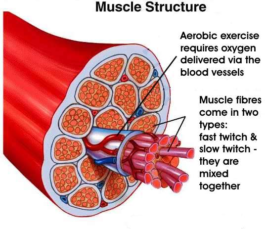
Diagram of neuromuscular junction

11 of 30

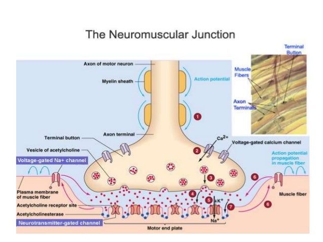
Diagram 2: Neuromuscular Junction

12 of 30

Nerve Impulse at Neuromuscular Junction

  • reaches synaptic knob
  • synaptic vesicles fuse with presynaptic membrane
  • releases acetylcholine into synaptic cleft
  • attaches to receptor sites on postsynaptic membrane = membrane of muscle fibre
  • alters permeability to Na+ ions - enter rapidly = DEPOLARISATION
  • neurotransmitter = acetylcholine
  • broken down by acetylcholinerase
  • muscle isn't over-stimulated
  • choline + ethanoic (acetyl) acid diffuses back into neurone
  • recombine to form acetylcholine
  • energy (ATP) supplied by mitochondria
13 of 30

Transmission Electron Microscopy (TEM) of skeletal

14 of 30

Neuromuscular Junction vs. Cholonergic Synapse

Similarities:

  • neurotransmitters are transported by diffusion
  • receptors - bind with neurotransmitter - influx of Na+
  • use Na+/K+ pump to repolarise axon
  • enzymes break down neurotransmitter
15 of 30

Neuromuscular Junction vs. Cholonergic Synapse

Differences:

Neuromuscular Junction

  • excitatory
  • neurones link to muscles
  • motor neurones
  • end of action potential
  • acetylcholine binds to receptors on membrane of muscle fibre

Cholinergic Synapse

  • excitatory or inhibitory
  • neurones link to neurones or effectors
  • motor, sensory or intermediate (relay) neurones
  • new action potential - postsynaptic membrane
  • acetylcholine binds to receptors on postsynaptic membrane
16 of 30

Contraction of skeletal muscle

  • movement
  • muscles attached to skeleton
  • incompressible bone skeleton
  • muscle exerts force - bones move around joints
  • the muscle does not change shape
  • move part of skeleton
  • one muscle can't move part in opposite directions
  • another muscle = ANTAGONISTIC PAIR
  • pull in opposite directions
  • one contracts + one relaxes
17 of 30

Sliding filament mechanism

  • contraction of muscle fibre
  • actin + myosin filaments slide past one another
  • muscle function depends on shape of proteins

(http://img1.mnimgs.com/img/study_content/curr/1/11/14/227/3744/NS_15-10-08_Reena_11_Biology_20_10_JYO_html_m76a3fa81.jpg)

18 of 30

Evidence for the sliding filament mechanism

  • more overlap of actin + myosin during contraction

Changes to sarcomere:

  • I-band narrows
  • Z-lines are closer together = shorter sarcomere
  • H-zone narrows
  • A-band stays the same width - length of myosin
  • muscle contraction not due to shorter filaments
19 of 30

Myosin

2 proteins:

  • fibrous protein - filament - made up of lots of molecules (tail)
  • globular protein - two bulbous structures (head)

(http://legacy.owensboro.kctcs.edu/gcaplan/anat/images/Image325.gif)

20 of 30

Actin

  • globular protein
  • molecules in long + twisted chains
  • helical strand

TROPOMYOSIN

  • long + thin threads
  • wound around actin filaments

21 of 30

The sliding filament mechanism of muscle contracti

  • actin + myosin filaments slide past each other
  • changes in band patterns on myofibrils
  • bulbous heads of myosin form cross bridges with actin
  • attach to binding sites on actin
  • flex in unison - pull actin filaments along myosin filaments
  • ATP - source of energy
  • detach - return to original angle
  • re-attach further along the actin
  • continuous process
22 of 30

Muscle Stimulation

  • action potential reaches many neuromuscular junctions simultaneously
  • Ca2+ ion protein channels open
  • Ca2+ ion diffuse into synaptic knob
  • synaptic vesicles fuse with presynaptic membrane
  • release acetylcholine (neurotransmitter) into synaptic cleft
  • acetylcholine diffuses across synaptic cleft
  • binds with receptors on the muscle cell-surface membrane
  • DEPOLARISATION
23 of 30

Muscle Contraction

WAVE OF EXCITATION

  • action potential travels into fibre
  • via tubules (T-tubules) - extensions of cell-surface membrane
  • in cyctoplasm of muscle - sarcoplasm
  • tubules - ER of muscle = sarcoplasmic reticulum
  • active transport of Ca2+ ion from cytoplasm - low concentration
  • Ca2+ protein channels open
  • Ca2+ diffuse into muscle cytoplasm
  • down concentration gradient
  • Ca2+ ions cause tropomyosin to pull away 
  • Ca2+ ions change tertiary structure of tropomyosin - protein!
  • bindings site are no longer blocked
24 of 30

Muscle Contraction (cont.)

  • ADP attach to myosin head
  • binds to actin + forms a cross bridge
  • attached to actin - myosin heads change angle
  • pulls actin along - releases ADP
  • ATP attaches to each myosin head
  • myosin heads detach from actin
  • Ca2+ ions activate ATPase - forms ADP + Pi via hydrolysis
  • provides energy for myosin to return to its original position
  • myosin head + ADP - reattaches further along actin
  • cyclical process - needs a high concentration of Ca2+ in the myofibril
25 of 30

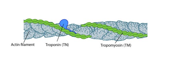
Diagram of Muscle Contraction

26 of 30

Muscle Contraction (cont.)

  • myosin are joined tail to tail
  • move in opposite directions
  • actin moves in opposite directions
  • actin filaments move towards each other
  • shorter distance between Z-lines
  • movement
27 of 30

Muscle Relaxation

  • no nervous stimulation
  • Ca2+ actively transported into endoplasmic reticulum
  • energy - hydrolysis of ATP to ADP + Pi
  • reabsorption of Ca2+ - tropomyosin moves back
  • binding sites are blocked again
  • myosin heads can't bind to actin
  • no muscle contraction
  • muscle relaxes
  • force from antagonistic muscles pulls actin out from between myosin
28 of 30

Energy supply during muscle contraction

  • needs lots of energy
  • hydrolysis of ATP to ADP + Pi
  • movement of myosin heads
  • reabsorption of Ca2+ ions into endoplasmic reticulum by active transport
  • escape from danger - rapid response - life-saving
  • most ATP is regeneratedrespiration of pyruvate in mitochondria
  • lots of mitochondria in muscle
29 of 30

Role of Phosphocreatine

ENERGY SUPPLY

  • demand for oxygen is greater than the rate of its supply
  • need for anaerobic respiration
  • PHOSPHOCREATINE + glycolysis
  • can't supply energy directly
  • regenerates ATP
  • stored in muscle
  • reserve supply of phosphate
  • ADP + Pi -> ATP
  • muscle relaxes - store of phosphocreatine is replenished - Pi from ATP 
30 of 30

Comments

No comments have yet been made

Similar Biology resources:

See all Biology resources »See all Muscles resources »