BY1 - Cell Structure and Organisation
- Created by: zopetre_
- Created on: 18-03-17 10:33
Animal cell structure

Plant cell structure

Describe the structure/role of the nucleus
The nucleus is usually spherical and 10-20um in diameter. It contains DNA, comprising of chromosomes which direct protein synthesis. DNA also provides a template for DNA replication.
The nucleus has many structures:
- Bounded by a double membrane, the nuclear envelope, which has pores within it to allow large molecules (mRNA and ribosomes) to leave the nucleus.
- Granular material, nucleoplasm, which contains chromatin.
- Small, spherical bodies each called a nucleolus which are the sites of formation of rRNA.

Describe the structure/role of mitochondria
Mitochondria are often cylindrical and 1-10um in length. The function of the mitochondria is to produce ATP during aerobic respiration. Metabolically active cells, such as muscle cells, contain many mitochondria due to the high metabolic activity taking place.
The mitochondria consists of many structures:
- Two membranes separated by a narrow, fluid-filled inter-membrane space. Inner membrane is also highly infolded to form cristae which provide a large surface area for the attachment of enzymes involved in respiration.
- Organic matrix which contains many compounds, including lipids and proteins.
- Small 70S ribosomes and a small circle of DNA, mitochondria can make their own proteins and also self-replicate
Describe the structure/role of chloroplasts
Chloroplasts occur in the cells of photosynthesising tissue. They are most likely found in the palisade mesophyll cells, below the upper surface of the leaf.
Each chloroplast is surrounded by two membranes, they have several other structures:
- A stroma which is fluid-filled and contains some of the products of photosynthesis, such as lipid droplets and starch grains.
- 70S ribosomes and circular DNA, enabling them to make their own proteins and self-replicate.
- Within the stroma are many closed, flattened sacs - thylakoids. A stack of thylakoids is called a granum. And each granum consists of between two and a hundred sacs. Photosynthetic pigments such as chlroophyll are found in the thlakoids. The arrangement products a large surface area.
Describe the structure/rule of endoplasmic reticul
Endoplasmic reticulum is an elborate system of parallel, double mrmbanes forming flattened sacs with interconnected, fluid-filled spaces between them, cisternae. It's conected with the nuclear envelope, allowing transport of materials through the cell. There are two types:
- Rough ER has ribosomes on the outer surface membrane. it transports proteins made there. It is present in large amounts in cells that make a lot of protein, such as amylase in the salivary glands.
- Smooth ER consists of membranes that lack ribosomes. It's associated with the synthesis and transport of fatty acids and lipids.
Describe the structure/role of ribosomes
Ribosomes can exist in two different sizes, 70S ribosomes are smaller and found in prokaryotic cells, whereas, 80S ribosomes are larger and found in eukaryotic cells. They are found freely in the cytoplasm of prokayotic cells, and on the RER in eukaryotic cells.
They have one large and one small subunit. They are assembled in the nucleolus from rRNA and protein. They are important in protein synthesis as they're the site of translation.
Describe the structure/role of the Golgi body
Golgi body resembles the structure of ER, but it more compact. It is formed of vesicles which contain polypeptides, pinching off from the RER and fusing with the stack of membranes. Proteins are modified and packaged. At the other end, vesicles containing modified proteins are pinched off. Vesicles carry proteins elsewhere to the cell.
The Golgi Body has many functions: producing secretory enzymes, secreting carbohydrates, producing glycoprotein, transporting/storing lipids and forming lysosomes.

Describe the structure/role of lysosomes
Lysosomes are small, temporary vacuoles surrounded by a single membrane. They are formed when they are pinched off from the Golgi body.
They contain and isolate potentially harmful digestive enzymes from the rest of the cell, and release these enzymes when the cell needs to recycle worn out organelles. The enzymes can also digest material that's been taken into the cell.
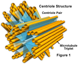
Describe the structure/role of centrioles
Centrioles occur in animal cells and most protoctistans. They're located outside of the nucleus.
They are two rings of microtubules which make hollow cylinders positioned at right angles to eachother.They may often be referred to as the centrosome. They are important in cell division, where they organise the microtubules that make the spindle.
Describe the structure/role of the vacuole
The vacuole is a large permanent structure in plant cells. It consists of a fluid-filled sac which is bounded by a single membrane called the tonoplast. They contain cell sap, which is a solution that stores chemicals such as glucose. They have a major role in supporting soft plant tissues.
Differences between plant and animal cells
Describe prokaryotic cells
Prokaryotic cells are single-celled organisms which lack membrane-bound organelles. They have no nucleus or any internal membranes. An example of a prokaryotic cell is bacterium.
All prokaryotes have: DNA molecules loose in the cytoplasm, a peptidoglycan cell wall, 70S ribosomes, cytoplasm and a cell membrane.
Only some prokaryotes have: a slime coat, flagella, photosynthetic lamellae holding photosynthetic pigments, mesosome and plasmids.
Compare eukaryotic and prokaryotic cells
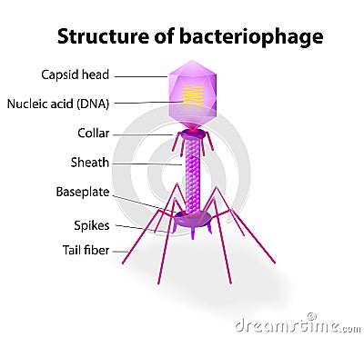
Describe the structure/role of viruses
Viruses are extremely small and can only be seen with an electron microcope. They aren't made of cells so are acellular. There are no organelles, choromosomes and no cytoplasm. It exists as a inert virion outside of a living cell. They are made up of a core of nucleic acid, DNA or RNA, surrounded by a protein coat called the capsid.
When they invade a cell they are able to take over the cells metabolism and multiply inside the host cell. The viruses that attack bacteria are called bacteriophages. The only characteristic of life that viruses display is their ability to reproduce.
Describe epithelial tissue
Epithelial tissue forms a continuous layer, covering/lining the internal and external surfaces of the body. They have no blood vessels, may have nereve endings and cells sit on a basement membrane made of colagen/protein, they vary in shape and complexity. They ofen have a protective/secretory function.
Cuboidal epithelium is the simplest form, cells have a cube shape and tissue is one cell thick. It occurs in the proximal convoluted tubule of the kidney nephron or ducts of sailvary glands.
Columnar epithelium have elongated cells. They have cilia. They're found in the oviduct and trachea.
Squamous epithelium constis of flattened cells on a basement membrane. Form the walls of the alveoli, and line the renal capsule of the nephron.
Describe muscle tissue
It comes in three main types, with different structures and functions:
- Skeletal muslce is attached to bones, generates locomotion in mammals. Bands of long cells/fibres, giving powerful contractions. Muscles tire easily. Voluntary muscles. Striated muscle.
- Smooth muscle has indiviudal spindle-shaped cells, contract rhythmically but less powerfully than skeletal. Occur in the skin, walls of blood vessels and digestive/respiratory tracts. Involuntary muscles. Unstriated muscle.
- Cardiac muscle is only found in the heart. Structures and properties are between skeletal and smooth muscle. Cells are striated, but lack long fibres of skeletal muscle. They contract rhythmically. It doesn't tire.

Comments
No comments have yet been made