Biological Membranes

?
  • Created by: rosieevie
  • Created on: 14-01-17 21:01

Importance of Membrane Proteins

  • 20% human genome codes for membrane proteins
  • Important interface of cell
  • Dictate how cell reacts with environment
  • Important drug targets
  • Faulty membrane proteins cause:
    • Cystic fibrosis
    • Malignant hypothermia
1 of 14

Eukaryotic Cell Membranes

  • Internal organelles bounded by membranes
  • Membranes invisible to light microscopes (okay with electron)
  • Two major components:
    • Lipids -small non-polar soluble molecules = permeability barrier
    • Proteins = biological properties e.g. transport, signalling, adhesion
2 of 14

Membrane Lipids - Phospholipids

  • Glycerol-based molecules e.g. phosphatidycholine

(http://nootropicsupplementreview.com/wp-content/uploads/2014/05/phosphatidylcholine.png)

  • Sphinogosine-base molecules e.g. sphingomyelin(http://aocs.files.cms-plus.com/LipidsLibrary/images/ImportedImages/lipidlibrary/Lipids/sph/Figure1.png)
  • Also contain sterols e.g. cholesterol 

(http://2.bp.blogspot.com/-rZ9GgpNvt6A/USY70k2fJGI/AAAAAAAABUU/VU1JbsXljXQ/s400/Cholesterol.gif)

3 of 14

Membrane Lipids

  • Ampipathic - contain polar and non-polar groups
  • In an aq. environment hydrophobic effect occurs:
    • Driven by water 
    • Hydrophobic mol. interfere with water's hydrogen bonds
    • Water will squeeze lipids together to minimise interactions of hydrophobic areas to water
    • Spontaneous formation of lipid bilayers (act as permeability barriers for polar/large mols) or self-sealing vesicles in water 
  • Membranes not spontaneously produced - only build from inserting lipids into new bilayers
  • Enzymes move phospholipids to either side - flippases (to cytoplasmic side) and floppases (to extracellular fluid) = transverse diffusion
4 of 14

Liquid Crystalline Phase

  • Membranes have lots of lipids with acy chains containing cis-double bonds
  • Cis-bonds prevent close acyl chain packing = motile
  • Liquid crystaline phase = lipids can move around in bilayer (low melting temperatures)
  • This is called lateral diffusion - same side
  • Essential for function at low temperatures 
  • Low low temperatures = gel phase and acyl chains frozen - lipids cannot move
  • Membrane proteins must undergo conformational changes and interact with adjacent proteins
5 of 14

Membrane Proteins

  • Provide control for the internal cell/organelle environment
  • Transporters, channels, receptors
  • All can be transmembranous
  • a-helical conformation (stabilised by H bonds) means they can cross hydrophobic core
  • Transmembrane domain residues are hydrophobic
  • End residues are polar
  • R-groups project outwards = anchorage in bilyaer
  • Single helix cannot form route for polar solutes as they are too big/polar
  • Amphipathic helicases (~4) cluster w/ polar resiudes on inside = route
6 of 14

Gated Ion Channels

  • Makes membrane selective
  • Charge at beginning of channel = only opposite charge attracted
  • Gate required to stop flow of ions (depending on electrochemical gradient)
  • Helicases rotate to expose small polar groups and open channel
  • When gate closed hydrophobic groups on inside = no polar molecules
  • Ligand binds to cause helicases to rotate
  • Cannot
    • Move ions/mols against concentration gradient
    • Facilitate large molecules (ions would sneak through, disrupting conc. gradients)
7 of 14

Transporters

  • 2 gates - neither open at same time = no major change in ion gradients
  • Solute recognition site - solute binds causing 1st gate to shut and 2nd to open
8 of 14

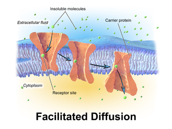
Facilitated Diffusion

  • Transporter undergoes conformational change e.g. rocking
  • Molecule binds tightly in either direction
  • No net transport after equilibrium 
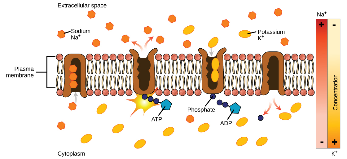
9 of 14

Active Transport

  • Against a concentration gradient
  • Molcule needs to bind tightly then let go at end
  • Requires ATP to overcome attraction force = pull molecule and channel apart
10 of 14

Modelling Membrane Proteins

  • Do not readily form 3D structures = few been mapped
  • Difficult to disrupt bilayer and isolate proteins
  • Most models based on sequence analysis and indirect studies
  • You need to form protein crystals and fire x-ray beans to be scattered = diffraction pattern
  • Must trap proteins in various conformations = flip-book method

Labelling Studies

  • Identify surface exposed proteins using membrane impermeant reagents - covalent modifers modifying specific residues on outside = analyse sequence
  • Identify transmembrane reagents using hydrophobic labelling reagents
    • Must first break membrane to allow reagent to dissolve in bilayer 
    • Use detergent = collapse and micelles form
    • Proteins react with detergent = protected in shape
    • Can be seperated and purified
11 of 14

Modelling Membrane Proteins - Sequence Analysis

  • Reveal potential transmembrane helices 
  • Amphipathic helicases hard - polar amino acids break up line of non-polar ones
  • Hydropathy Plots:
    • Each amino acid assigned a value for its hydrophobicity (energy required to move residue from aqueous to inside bilayer)
    • Higher value = more hydrophobic 
    • Average length of transmembrane helix 20 residues -> look for 20 consecutive hydrophobic amino acids
    • Computer plots = potential transmembrane helicases in peaks
    • No idea of orientation of proteins 
    • Combo of this and labelling studies required
12 of 14

Glycosylation of Membrane Proteins

  • Glycophorin A etc. are glycosylated on extra cellular surface
  • Precise pattern depends on battery of inherited glycosylation enzymes of an individiual
  • Gives rise to ABO blood group system
13 of 14

B-Barrel Structures

  • Less common
  • Formed of antiparralell B-sheets -> barrel structure
  • Often got alternating hydrophobic and philic residues (polar inside and non-polar outside = hydrophilic pore/porin
  • Not found in plasma membrane - ion concentration gradient issues
  • Found in mitochondria
    • Porous membrane preserves intermembrane space = required for mitochondria function
  • Found in outer membranes of gram negative bacteria e.g. E.coli
    • Live in hostile environmnets with protases
    • Protects the inner layer which contains transporters
    • Outer layer allows mols. to enter periplamic space down conc gradient
    • Prevention of proteases entering
14 of 14

Comments

No comments have yet been made

Similar zoology resources:

See all zoology resources »See all Membrane Proteins resources »